Pancreatic PEComa


Pancreatic perivascular epithelioid cell tumors (or "Pancreatic PEComas") are a subtype of the larger family of PEComas. Pancreatic PEComas are very rare with <20 cases described.
Clinical presentation
More common in adults (in contrast to lymphangiomas in the head and neck, which are more common in children). Account for approximately 1% of abdominal lymphangiomas. More common in women
Pathology
As their name suggests, these tumors arise from perivascular epithelioid cells and occur due to a genetic alteration in the tuberous sclerosis gene complex. The PEComas are a group of tumors, including
- clear cell "sugar" tumors
- angiomyolipoma
- lymphangiomyomatosis
The cells are periodic acid-Schiff stain (PAS) positive and diastases sensitive. Immunohistochemistry markers include: smooth muscle and melanocytic markers (SMA, HMB-45, and HMSA-1)
Radiographic features
CT
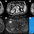
- tend to occur in the head and body of the pancreas
- well-demarcated
- hypoenhancing mass with a hypervascular capsule
- may demonstrate hemorrhage or cystic degeneration
- fat attenuation may be present
MRI
Signal characteristics include
- T1: hypointense
- T2: hyperintense
- T1 C+: heterogeneous enhancement
- T1 FS: macroscopic fat intensity may be present (saturates on a fat sat sequence)
Ultrasound
- nonspecific pancreatic mass
- heterogeneous echogenicity
- well-encapsulated
Treatment and prognosis
Given the rarity of the lesion, there is no standard treatment
Differential diagnosis
A pancreatic PEComa should only be considered if a pancreatic mass is well-encapsulated, but since it is so rare, it should never be at the top of a differential. It's probably only reasonably suggested in a patient with tuberous sclerosis and a pancreatic mass.
Other similar pancreatic masses that should be considered before it are
- pancreatic endocrine tumor ("islet cell" tumor)
- solid-pseudopapillary tumor (SPT)
- metastasis to the pancreas
- also rare, but much more likely than a pancreatic PEComa
Siehe auch:

 Assoziationen und Differentialdiagnosen zu perivaskulärer epitheloidzelliger Tumor (PECom) des Pankreas:


