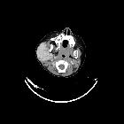
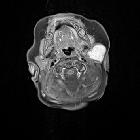
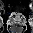
Parotid infantile hemangioma

Imaging of
parotid anomalies in infants and children. Infantile hemangioma. Lobulated, hypoechoic and hypervascular mass (a) containing high velocity and low resistance arteries (b), on ultrasonography. Diffuse enlargement of the left gland by strongly enhanced mass containing tortuous vessels responsible for flow void images [white arrows], on axial T2-weighted image (c) and T1-weighted image, after intravenous injection of gadolinium chelate (d)

Parotid
infantile hemangioma • Infantile hemangioma - parotid - Ganzer Fall bei Radiopaedia

Parotid
infantile hemangioma • Parotid infantile hemangioma - Ganzer Fall bei Radiopaedia
Parotid infantile hemangiomas are the most common parotid tumor of childhood. They usually run a characteristically benign course.
Epidemiology
The median age at diagnosis is 4 months . There is a female preponderance with a male: female ratio of 1:3.
Clinical presentation
Presents as an enlarging parotid mass in an infant that was not present at birth. a cutaneous component (e.g. strawberry lesion) may be present. They can occasionally act as significant vascular shunts if they do not involute.
Radiographic features
Ultrasound
- homogeneous echogenic parotid mass
- lobulated
- fine echogenic internal septations
- replaces or expands parotid tissue
- large internal vessels
- rarely flow only seen on gray-scale as it is too low to be seen on Doppler
MRI
- T1: intermediate signal, between that of muscle and fat
- T2: hyperintense
- prominent flow voids
- T1 C+ (Gd): homogeneous enhancement
Treatment and prognosis
Surgery is usually avoided since there is a risk of facial nerve damage and most lesions resolve over time either spontaneously or with medical therapy .
Siehe auch:

 Assoziationen und Differentialdiagnosen zu infantiles Hämangiom der Glandula parotis:
