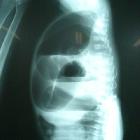
Pediatric abdomen (prone cross-table lateral view)

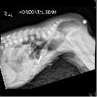

The prone cross-table lateral view is an additional projection to demonstrate the pediatric abdomen and is a more ideal alternative to the invertogram, which may be less comfortable for the patient. This discomfort may result in a continuously crying baby, causing the puborectalis sling to contract, leading to a misleading impression of distal rectal obscuration .
Indications
This view is ideal for indicating the distance between the gas bubble in the terminal colon and the perineal skin, allowing the classification of anal atresia in neonates. The image is often obtained 24 hours after birth, to allow for small fistulas to become apparent.
Patient position
- patient is prone in genupectoral position (for a minimum of 3 minutes)
- ensure no rotation of hips and shoulders
- remove any radiopaque items (e.g. ECG dots, diaper, shiny decorative clothing)
- take the x-ray in full inspiration
- a radio-opaque marker (i.e. a coin) is placed over the expected anus using radiolucent tape
Technical factors
- lateral projection
- suspended inspiration (on observation)
- centering point
- the midcoronal plane at the level of the greater trochanter
- collimation
- superior to the diaphragm
- inferior to the rectum
- anteroposterior to include soft tissue edge
- orientation
- landscape
- detector size
- will vary depending on the child's body habitus
- exposure
- 60-75 kVp
- 3-10 mAs
- SID
- 100 cm
- grid
- if patient thickness is above 10 cm, use of a grid is advisable
Image technical evaluation
- it should be possible to determine the distance between the air-filled distal rectal pouch and the anal dimple (marked by a radio-opaque marker)
- the abdomen should be free from rotation
- no blurring of the bowel gas from respiratory motion is ideal
Practical points
Preparing the room beforehand (setting up the detector, exposure and preparing lead gowns) is extremely beneficial as patients may get startled by the movement of loud equipment.
Imaging of neonates whilst they are calm or asleep can allow for a less challenging examination.
Immobilization techniques
To prevent malrotation, motion artefact in the radiograph, parental holding at the head and leg of the patient may be required. Placing immobilization devices to ensure the patient is faced down with their hips flexed will allow for increased stability in positioning.
Siehe auch:


 Assoziationen und Differentialdiagnosen zu Pediatric abdomen (prone cross-table lateral view):