anal atresia


 nicht verwechseln mit: Morbus Hirschsprung
Anal atresia, or imperforate anus, refers to a spectrum of anorectal abnormalities ranging from a membranous separation to complete absence of the anus.
Epidemiology
The estimated incidence is 1 in 5000 live births.
Pathology
Clinically there is no anal opening. Subtypes can be classified into two broad categories: high (supralevator) or low (infralevator), depending on the location of the atretic portion.
Genetics
Most cases are sporadic, with occasional familial forms
Associations
There are frequent associations with other congenital abnormalities.
- other atresias
- VACTERL association
- caudal regression syndrome: associated sacral agenesis and lower limb hypoplasia
- Currarino's triad; anorectal malformations with sacral anomalies and presacral mass lesion
- fistulous tracts to the urethra or vagina may be present or may have a single cloacal opening
Radiographic features
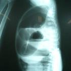
Abdominal radiograph
- can be variable depending on the site of atresia (i.e. high or low), level of meconium impaction and physiological effects such as straining
- may show multiple dilated bowel loops with an absence of rectal gas
air within urinary bladder suggests high type
- calcified meconium in the bowel loops would suggest high type (meconium calcifies due to urine exposure)
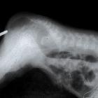
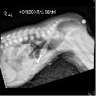
Invertogram
A coin/metal piece is placed over the expected anus and the baby is turned upside down (for a minimum 3 minutes).
The distance of the gas bubble in the rectum from the metal piece is noted:
- >2 cm denotes high type
- <2 cm denotes low type
False-positive: if image is taken in the 1st 24 hours of life or impacted meconium in distal rectum .
For radiographic technique, see invertogram view and prone cross-table lateral view articles.
Fluoroscopy (contrast study)
- to detect a rectourinary, rectovaginal, or rectoperineal fistula
- the fistula is considered low (below the levator ani plane) if it is below the pubococcygeal line and high if above it
Ultrasound
- the anus may be seen as an echogenic spot at the level of the perineum and in anal atresia, this echogenic spot may be absent
- may show bowel dilatation
- an infracoccygeal or transperineal approach may allow differentiation between high and low subtypes
- kidneys should be assessed in such patients
- spinal US can reveal spinal cord lesions like tethering of cord
MRI
Can be used pre/postoperatively to study pelvic floor, renal and spinal abnormalities .
Treatment and prognosis
- low subtypes are treated with anoplasty
- high subtypes are treated with colostomy with subsequent potential repair
Complications
See also
Siehe auch:
- caudal regression syndrome
- Ösophagusatresie
- VACTERL-Assoziation
- Invertogramm nach Wangensteen-Rice
- Duodenalatresie
- Mekoniumperitonitis
- Stratton-Parker-Syndrom
- small bowel atresia
und weiter:
- ASP-Assoziation
- Down-Syndrom
- Ileumatresie
- caudal dysplasia sequence
- VATER
- OEIS complex
- esophageal atresia with H-type tracheo-esophageal fistula
- Pallister-Hall-Syndrom
- VATER anomaly
- Atresie
- fetal bowel dilatation
- IVIC-Syndrom
- H-type tracheo-oesophageal fistula with no atresia
- McKusick-Kaufman-Syndrom
- fetal colonic dilatation
- angeborene ösophagotracheale Fistel
- Schmid-Fraccaro-Syndrom
- Kolonatresie
- antenatale Darmperforation
- anorektale Stenosen


 Assoziationen und Differentialdiagnosen zu Analatresie:


