perianal fistula


Perianal fistula (or fistula-in-ano) (plural: fistulae or fistulas) is the presence of a fistulous tract across/between/adjacent to the anal sphincters and is usually an inflammatory condition .
Epidemiology
Incidence is estimated at ~1:10,000 , with a recognized male predilection of 2-4:1.
Associations
- spontaneous
- diverticulitis: perhaps the most common cause in developed countries
- Crohn disease
- it is unclear if ulcerative colitis is a risk factor for perianal disease
- other bowel or pelvic infections
- tuberculosis
- iatrogenic: post-surgical complication
- post-ultralow anterior resection
- hysterectomy
- post-pelvic radiotherapy
- childbirth
- pelvic malignancies
Clinical presentation
Symptoms are variable, including anal pain, tenesmus, pruritus, and sepsis.
Pathology
The most commonly accepted pathophysiology is the cryptoglandular hypothesis, which suggests that obstruction of the deep submucosal glands results in infection and abscess formation in the inter-sphincteric space, which consequently drains inferiorly between the sphincters, opening onto the skin surface or, less commonly, erodes through both the internal and external sphincters into the ischiorectal space, then onto the skin surface .
Transsphincteric fistulae are secondary to ischiorectal abscesses, with a resultant extension of the tract through the external sphincter. Intersphincteric fistulae are due to perianal abscesses. Suprasphincteric fistulae are due to supra levator abscesses.
Location
Goodsall's rule states that the internal opening of the fistula is dependent on where the fistula is located relative to the 'anal clock' (i.e. with the patient in the lithotomy position, anterior is 12 o'clock and posterior is 6 o'clock) and the transverse anal line (a line drawn from 9 o'clock to 3 o'clock):
- if the internal opening is anterior to the transverse anal line there will be a (usually simple) direct radial fistulous tract
- if the internal opening is posterior to the transverse anal line there will be a tortuous (and often more complex) fistulous tract that enters posteriorly in the midline (6 o'clock)
Classification
Surgical classification
The Parks classification has become the most widely used surgical classification for distinguishing four types of fistula. The course of the fistula and its relationship to the anal sphincters is described in the coronal plane :
- intersphincteric (~70%): fistula crosses the intersphincteric space and does not cross the external sphincter
- transsphincteric (25%): fistula crosses from the intersphincteric space, through the external sphincter, and into the ischiorectal fossa
- suprasphincteric (5%): fistula passes superiorly into the intersphincteric space, and over the top of the puborectalis muscle then descending through the iliococcygeus muscle into the ischiorectal fossa and then skin
- extrasphincteric (1%): fistula crosses from the perineal skin through the ischiorectal fossa and levator ani muscle complex into the rectum (i.e. it is outside the external anal sphincter)
Radiological classification
Radiologists have developed another grading system for perianal fistulae, which is based on landmarks on the axial plane and incorporates abscesses and secondary extensions to the grading system, and is called the St James’s University Hospital classification :
- grade 1: simple linear intersphincteric
- grade 2: intersphincteric with abscess or secondary tract
- grade 3: transsphincteric
- grade 4: transsphincteric with abscess or secondary tract within the ischiorectal fossa
- grade 5: supralevator and translevator extension
Radiographic features
Fistulography
Fistulography is a traditional radiological technique used to define the anatomy of fistulas, yet it is an unreliable technique and is difficult to interpret .
In fistulography, the external opening is catheterized with a fine cannula, and a water-soluble contrast agent is injected to define the fistulous tract .
It has two major drawbacks :
- difficult to assess secondary extensions secondary to lack of proper filling with contrast material
- inability to visualize the anal sphincters and to determine their relationship to the fistula
Ultrasound
The benefits of ultrasonography over MRI are the former's ubiquity and lower operating costs . There are three ultrasonography methods for the evaluation of perianal fistulae, whether cryptoglandular or Crohn disease-associated :
- endoanal ultrasound (EAUS)
- transvaginal ultrasound (TVUS)
- transperineal ultrasound (TPUS)
These can also be applied in combination. Infusion of hydrogen peroxide into the fistulous tract renders it hyperechoic, thus facilitating its delineation. Doppler interrogation can show hyperemia in active Crohn disease.
Endoanal ultrasonography is deemed less sensitive than endoanal MR for deep supra levator disease .
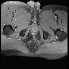
MRI
MRI is the imaging modality of choice. See pelvic MRI protocol for anal canal fistula assessment.
Active fistulous tracts are typically:
- T1: isointense to muscle
- T2: high signal compared to fat
- T2-FS: high signal compared to fat
- T1 C+: enhancing
Old, healed fistulae typically demonstrate low T1 and T2 signals without contrast enhancement, reflecting fibrosis.
Radiology report
- detection of the primary fistulous tract and its activity:
- active tract has high T2 signal and demonstrates intense enhancement
- chronic tracts have low signal on both T1- and T2-WI and will not show contrast enhancement
- location (right/left) and course
- relationship to the sphincter complex
- Parks classification: trans-, inter-, supra-, or extra-sphincteric
- the distance of the internal mucosal defect to the perianal skin on coronal images
- position of the internal mucosal opening on axial images
- use the "anal clock": anterior = 12 o'clock
- identify secondary fistulous tracts and the sites of any abscess cavities in order to avoid therapeutic failure and recurrence
- cranial extension above the levator ani muscle
Treatment and prognosis
The majority of perianal fistulas related to Crohn disease will not heal spontaneously and require surgical management.
Differential diagnosis
Siehe auch:
und weiter:


 Assoziationen und Differentialdiagnosen zu Analfistel:
