periventricular leukomalacia


Periventricular leukomalacia (PVL), or white matter injury of prematurity affecting the periventricular zones, typically results in cavitation and periventricular cyst formation.
It is important to note that both periventricular and subcortical leukomalacia corresponds to a continuous disease spectrum. Please refer to the article on patterns of neonatal hypoxic-ischemic brain injury for a relation between perinatal brain maturation process and these lesions.
Epidemiology
PVL is most common in premature neonates (less than 34 weeks gestational age with a median gestational age of 30 weeks) and <1500 grams at birth.
Clinical presentation
PVL may manifest as cerebral palsy (>50% in the setting of cystic PVL), intellectual disability or visual disturbance.
Pathology
It likely occurs as a result of hypoxic-ischemic lesions resulting from impaired perfusion at the watershed areas, which in premature infants are located in a periventricular location. It is likely that infection or vasculitis also play a role in pathogenesis.
- early: periventricular white matter necrosis
- subacute: cyst formation
- late: parenchymal loss and enlargement of the ventricles
Distribution
The white matter necrosis often occurs in a characteristic distribution with the pattern being dorsal and lateral to the lateral ventricles and with the involvement of the centrum semiovale, the optic (trigone and occipital horns) and acoustic (temporal horn) radiations.
Radiographic features
Ultrasound
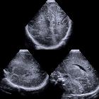
Cranial ultrasound provides a convenient, non-invasive, relatively low-cost screening examination of the haemodynamically-unstable neonate at the bedside. The examination also imparts no radiation exposure. Sonography is sensitive for the detection of hemorrhage, periventricular leukomalacia, and hydrocephalus.
On ultrasound, hyperechoic areas are firstly identified in a distinctive fashion in the periventricular area, more often at the peritrigonal area and in an area anterior and lateral to the frontal horns (periventricular white matter should be less echogenic than the choroid plexus).
These are watershed areas that are sensitive to ischemic injury. Follow-up scans in the more severely affected patients may reveal the development of cysts in these areas, known as cystic PVL (when cystic PVL is present, it is considered the most predictive sonographic marker for cerebral palsy).
Classification
See sonographic grading of PVL.
MRI
Initial MR images depict areas of T1 hyperintensity within larger areas of T2 hyperintensity.
Subsequent cavitation and periventricular cyst formation, features that are required for a definitive diagnosis of PVL, develop 2-6 weeks after injury and are easily seen on sonograms as localized anechoic or hypoechoic lesions. Progressive necrosis of the periventricular tissue with resulting enlargement of the ventricles is called end-stage PVL. CT and MR imaging findings of end-stage PVL include ventriculomegaly with irregular margins of the bodies and trigones of the lateral ventricles, loss of periventricular white matter with increased T2 signal, and thinning of the corpus callosum.
Differential diagnosis
The differential for periventricular echogenicity in neonates on ultrasound include
- cerebral edema: from various underlying causes
- cerebral infection: e.g. TORCH infection
- subependymal cysts
- choroid plexus cysts
- porencephaly
Siehe auch:
- Hirnödem
- subcortical arteriosclerotic encephalopathy (SAE)
- periventrikuläres Marklager SAE
- periventricular leukomalacia classification
- erweiterte Ventrikel
- neonatale hypoxisch-ischäme Enzephalopathie
- subkortikale Leukomalazie
und weiter:


 Assoziationen und Differentialdiagnosen zu periventrikuläre Leukomalazie:

