polyostotische fibröse Dysplasie

Fibröse
Dysplasie im Kalkaneus bei einem Patienten mit bekannter polyostotischer fibröser Dysplasie. Typische Darstellung im Röntgenbild mit milchiger, strukturloser Verdichtung.

Polyostotische
Fibröse Dysplasi. Hier Anteile im Kalkaneus, Kuboid und der Fibula. Oben Röntgen und CT. Unten MRT T1 coronar und PDW FS sagittal.
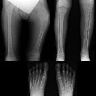
School ager
with left leg pain. AP radiograph of the femur (above left) shows a metaphyseal lesion that is expansile, lytic in appearance, with a narrow zone of transition and no periosteal new bone that has the appearance of a shepherd’s crook. AP radiograph of the bilateral tibia and fibula (upper right) shows multiple left tibial metaphyseal and diaphyseal lesions that are lytic in appearance with a narrow zone of transition and no periosteal reaction. AP radiograph of the feet (below) shows lesions in the diaphysis of the left first metatarsal and proximal phalanx that are lytic in appearance with a narrow zone of transition and no periosteal new bone.The diagnosis was polyostotic fibrous dysplasia.

Fibrous
dysplasia for radiologists: beyond ground glass bone matrix. The significance of MRI with diffusion-weighted imaging (DWI) in the patient with polyostotic fibrous dysplasia (FD). CT (a), T2-weighted MRI (b) and DWI (c) show multiple rib lesions (green arrows). The left rib lesion (red arrow) demonstrates restricted diffusion, which requires further evaluation to rule out malignant transformation

Fibrous
dysplasia for radiologists: beyond ground glass bone matrix. Nuclear medicine imaging in fibrous dysplasia (FD). a Bone scans with 99m-Tc-MDP are exquisitely sensitive at detecting the presence and extend of the disease. b 18-F-NaF on the same patient with polyostotic FD demonstrates multiple areas of focal radiotracer uptake corresponding to FD lesions. c, d 18-F-NaF PET/CT shows heterogeneous uptake by the lesions in the jaw and the spine with a central photopenic area (green arrow) and a peripheral metabolically active area (red arrow)

Fibrous
dysplasia for radiologists: beyond ground glass bone matrix. Multimodality imaging in polyostotic fibrous dysplasia (FD). a, b PET/CT with 18-F-NaF demonstrates multiple lucent FD lesions seen on CT with corresponding areas of mild radiotracer uptake on PET. c–f Lesions demonstrate intermediate T1 signal intensity on MRI (c), intermediate-to-low signal intensity on T2 (d), slightly hyperintense signal intensity on DWI (e), uniform enhancement after contrast administration (f)

Fibrous
dysplasia for radiologists: beyond ground glass bone matrix. Fibrous dysplasia (FD) of the spine. a Monostotic FD of the T6 vertebral body complicated by a compression fracture (green arrow). b Polyostotic FD involving the spine, ribs and skull (red arrows). c, d Postsurgical radiographs after a spinal fusion for scoliosis in the settings of polyostotic FD
polyostotische fibröse Dysplasie
Siehe auch:
- Fibröse Dysplasie
- McCune-Albright Syndrom
- polyostotic craniofacial fibrous dysplasia in infancy
- monostotische fibröse Dysplasie
- polyostotische kraniofaziale fibröse Dysplasie
und weiter:


 Assoziationen und Differentialdiagnosen zu polyostotische fibröse Dysplasie:
