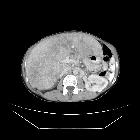
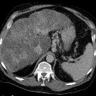
Pseudocirrhosis


Pseudocirrhosis is a radiological term used to recapitulate imaging findings of cirrhosis, but occurring in the setting of hepatic metastases. It is most commonly reported following chemotherapeutic treatment of breast cancer metastases, although has also been reported before treatment, and with other malignancies. It was, however, first described in tertiary syphilis.
Terminology
The term "pseudocirrhosis" is intended to convey a different disease process compared with cirrhosis of chronic liver disease. More rarely the term Hepar lobatum (carcinomatosum) is used.
Clinical presentation
Although most cases are mainly identified on an imaging basis, particularly found on post-treatment surveillance scans, patients may have symptoms related to liver failure and portal hypertension, including abdominal distension due to ascites.
Pathology
Two pathophysiologic correlates of pseudocirrhosis have been described :
- hepatic capsular retraction in response to chemotherapeutic agents, with nodular regenerative hyperplasia and absence of bridging fibrosis or cirrhosis on histopathology
- extensive fibrosis representing a profound desmoplastic response to an infiltrating tumor, occurring prior to chemotherapy
Radiographic features
Radiological manifestations on CT, ultrasound, and MRI may be identical to liver cirrhosis, and consist of:
- variable capsular retraction and liver nodularity
- hepatic segmental volume loss
- caudate lobe enlargement
- nodular contour
- confluent fibrosis
Siehe auch:

 Assoziationen und Differentialdiagnosen zu Pseudozirrhose der Leber:


