pulvinar sign

The pulvinar
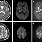
sign: T1-weighted sagital (A) and axial (B) MRI sections showing the pulvinar sign in a 66 year-old male patient. T1-weighted sagital (C) and axial (D) MRI section showing symmetrical high signals in the pulvinar region in a 42-year-old male patient. Courtesy: Dr Robert CARLIER and Dr Frédéric COLAS, CHU Raymond Poincaré, Garches, France.


 Assoziationen und Differentialdiagnosen zu pulvinar sign: