pyogenic cervicothoracic spondylodiscitis

Bone up on
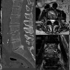
spinal osseous lesions: a case review series. Pyogenic spondylodiskitis. Sagittal STIR image (a) demonstrates abnormal marrow signal of C2, C3, C5, C6, and C7 (asterisks) with diffuse paravertebral soft tissue edema. Post-contrast T1-weighted image (b) demonstrates corresponding marrow enhancement, disc enhancement, and endplate erosion (orange arrows). Phlegmon or abscesses are seen at the level of C2-C3, C5-C6, and C6-C7 (white arrows), as well as epidural phlegmon extending along the dorsal aspects of C2 to C7. Sagittal T2 image (c) demonstrates fluid signal within the disc at C2-C3 and C5-C6 (black arrows), characteristic of pyogenic spondylodiskitis


 Assoziationen und Differentialdiagnosen zu pyogenic cervicothoracic spondylodiscitis: