Pyosalpinx


Pyosalpinx refers to a Fallopian tube that is filled, and often distended, with pus.
Pathology
A pyosalpinx often tends to be a complication of background pelvic inflammatory disease (PID). Inflammation results in tubal and peritubal adhesions with superimposed obstruction of the fimbrial end. Pyosalpinx can be a component of a tubo-ovarian abscess. Being unable to drain, the fallopian tube distends with pus, resulting in a pyosalpinx.
Radiographic features
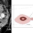
A pyosalpinx may be seen as a thickened fallopian tube and may or may not be associated with debris. The Fallopian tube may be distended.
Ultrasound
- dilated serpentine/tubular structure in the pelvis
- low-level echoes due to the higher protein content of the debris within the tube distinguish a pyosalpinx from a hydrosalpinx
MRI
Morphological appearances of a pyosalpinx can sometimes be indistinct from simple uncomplicated tubal dilatation (hydrosalpinx) . As opposed to a simple hydrosalpinx, in pyosalpinx the tube wall may appear thickened and have hyperenhancing tubal walls with surrounding inflammation.
Signal characteristics within the tubular lumen have been described as
- T1: variable due to varying protein content
- T2: often hyperintense, with characteristic amorphous shading
- T1 C+ (Gd): thick rim enhancement
- DWI/ADC: restricted diffusion in the fallopian tube
See also
Siehe auch:
und weiter:


 Assoziationen und Differentialdiagnosen zu Pyosalpinx: