Tuboovarialabszess


Tubo-ovarian abscesses are one of the late complications of pelvic inflammatory disease.
Epidemiology
Risk factors
Risk factors for tubo-ovarian abscesses include :
- previous pelvic inflammatory disease
- intrauterine device
- multiple sexual partners
- diabetes mellitus
- immunosuppression
- history of uterine surgery (e.g. post hysterectomy)
Clinical presentation
Patients typically present with a combination of fever, elevated inflammatory markers, lower abdominopelvic pain, and vaginal discharge. Fever and leukocytosis may sometimes be absent.
Pathology
Abscesses are often polymicrobial with a preponderance of anaerobic organisms .
Uncommon causes include actinomycosis, tuberculosis, and xanthogranulomatous inflammation .
Radiographic features
The clinical context is extremely important in radiological interpretation. Patients will experience tenderness with endovaginal scanning. Some differentiate between:
- tubo-ovarian "abscess": ovary and tube cannot be separately distinguished within the inflammatory mass
- tubo-ovarian "complex": if the tube and ovary are separately discernible structures within the inflammatory mass
Plain radiograph
Features are non-specific and may include:
- soft tissue density mass
- loss of normal pelvic fat planes
- an adynamic ileus may be present
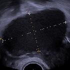
Ultrasound
Transabdominal and endovaginal ultrasound are the preferred initial imaging investigations. Findings may include:
- multilocular complex retro uterine/adnexal mass
- debris, septations, and irregular thick walls
- commonly bilateral
- echogenic debris within the pelvis
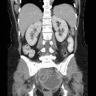
CT
Can be a helpful adjunct to ultrasound especially in determining the extent of disease :
- fluid attenuation pelvic masses which may contain fluid-fluid levels or gas
- usually shows a thick enhancing wall
- a tubular configuration is more conclusive of a pyosalpinx
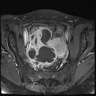
MRI
Can be useful especially when sonography is inconclusive or if the gas content is difficult to be differentiated from bowel gas .
Typically seen as thick-walled fluid-filled pelvic mass(es)
- T1: abscess contents typically hypointense
- T2: abscess contents typically heterogeneous signal or hyper-intense
Treatment and prognosis
Initial treatment can be with antibiotic therapy. Radiological guided drainage or surgery may be required in patients resistant to antibiotic treatment. Drainage may be performed from an endovaginal, transgluteal, or transabdominal approach, dependent on patient and operator preference .
Recognized complications include:
- abscess rupture
- rarely, perihepatitis (Fitz-Hugh-Curtis syndrome)
Differential diagnosis
Clinical features of infection is a key to aid diagnosis as a number of other pathologies can give similar appearances :
- pelvic malignancy
- complex diverticular abscess
- complex appendiceal abscess
- pelvic endometriosis
- pelvic hematoma
- pelvic hemorrhagic cysts
- hydrosalpinx
- ectopic pregnancy
- inflammatory bowel disease
- pelvic inflammatory disease
- ovarian torsion
- pyelonephritis
Uncommon causes of tubo-ovarian abscesses such as actinomyces and tuberculosis have many overlapping features with ovarian malignancy, including:
- relatively vague presentation
- solid/cystic ovarian masses
- peritoneal and/or serosal thickening and enhancement
There may be clues that favor uncommon causes of tubo-ovarian abscesses over malignancy:
- long-standing intrauterine contraceptive device as a risk factor for actinomyces
- smoother peritoneal enhancement more typical of peritonitis rather than carcinomatosis.
In these cases, biopsy or fluid sampling is often most appropriate to guide therapy and avoid unnecessary surgical intervention .
Siehe auch:
- Endometriose
- Unterleibsentzündung
- tuboovariale Tuberkulose
- Tuberkulose des Ovars
- infiziertes Teratom des Ovars
- infizierte Ovarialzyste
- pelvic inflammatory disease and tubo-ovarian abscess
und weiter:


 Assoziationen und Differentialdiagnosen zu Tuboovarialabszess: