Pyelonephritis
Pyelonephritis
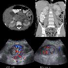
beidseits: Multiple, radiäre Hypodensitäten / Minderperfusionen in der Rinde beider Nieren. Klinisch Urosepsis. E. coli.

Akute
Pyelonephritis links mit Minderperfusion der linken Niere und deutlicher perirenaler Mitreaktion. Oben arterielle Phase axial und koronar, unten portalvenös.

Infected
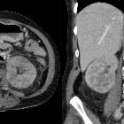
isolated aneurysm of the left common iliac artery. CE-MDCT - venous phase shows a globular kidney, mild hydronephrosis (blue arrow) and low-attenuation small cortical fluid collections (orange arrow) in keeping with complicated pyelonephritis.

Pyelonephritis
• Pyelonephritis - Ganzer Fall bei Radiopaedia

Correlation
of contrast-enhanced ultrasonography and computed tomography in complicated pyelonephritis. A low attenuating mass with low peripheral enhancement of the right kidney (red circle) was detected. Considering the clinical features of the patient, the findings implied complicated pyelonephritis.

Correlation
of contrast-enhanced ultrasonography and computed tomography in complicated pyelonephritis. In delayed phase, the perfusion of the lesion remained poor (red circle).

Correlation
of contrast-enhanced ultrasonography and computed tomography in complicated pyelonephritis. After the microbubbles intravenous administration, a low-perfusion lesion (red circle) was revealed.
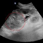
Correlation
of contrast-enhanced ultrasonography and computed tomography in complicated pyelonephritis. Enlargement of the right kidney and an upper pole hyperechoic mass (red circle) are visualized.

Horseshoe
kidney • Pyelonephritis in horseshoe kidney - Ganzer Fall bei Radiopaedia

Pyelonephritis
• Pyelonephritis in pregnancy (MRI) - Ganzer Fall bei Radiopaedia

Pyelonephritis
• Pyelonephritis - Ganzer Fall bei Radiopaedia

Infected
isolated aneurysm of the left common iliac artery. CE-MDCT - arterial phase shows hydroureter (blue arrows), the aneurysm (orange arrow), the high-crescent sign (green arrow) and the irregular outline of the contained rupture (white arrow).

Imaging for
acute pelvic pain in pregnancy. A 33-year-old woman at 32-gestation week was admitted manifesting fever and acute pelvic pain. Coronal diffusion-weighted (DW) image (a) and ADC map (b) show a focal area of restricted diffusion at the level of the upper pole of the left kidney, not seen at T2 Haste (c). The findings are indicative of focal pyelonephritis

Pyelonephritis
• Pyelonephritis - Ganzer Fall bei Radiopaedia

Chronic
pyelonephritis • Renal scarring - Ganzer Fall bei Radiopaedia

Emphysematous
pyelonephritis • Emphysematous pyelonephritis - Ganzer Fall bei Radiopaedia

Pneumobilia
• Acute pyelonephritis - Ganzer Fall bei Radiopaedia

Xanthogranulomatous
pyelonephritis • Xanthogranulomatous pyelonephritis - Ganzer Fall bei Radiopaedia

Renal
pseudotumor • Acute pyelonephritis - Ganzer Fall bei Radiopaedia

Pyelonephritis
• Acute bacterial pyleonephritis on MRI: value of DWI - Ganzer Fall bei Radiopaedia
Pyelonephritis (plural: pyelonephritides) refers to an upper urinary (renal) tract infection with associated renal pelvis, renal calyceal and renal parenchymal inflammation, and comprises a heterogeneous group of conditions.
- bacterial pyelonephritis
- chronic pyelonephritis
- renal tuberculosis
- emphysematous pyelitis
- emphysematous pyelonephritis
- malacoplakia
- fungal pyelonephritis
- xanthogranulomatous pyelonephritis (XGP)
See also
Siehe auch:
- Urolithiasis
- Niereninfarkt
- akute Pyelonephritis
- Nierentuberkulose
- xanthogranulomatöse Pyelonephritis
- Nierenabszess
- emphysematöse Pyelonephritis
- pyonephrosis
- perirenaler Abszess
- fleckige Nierenkontrastierung
- Pyelonephritis mit Nierenvenenthrombose
- Malakoplakie des Harntraktes
und weiter:


 Assoziationen und Differentialdiagnosen zu Pyelonephritis: