striated nephrogram


Striated nephrogram is a descriptive term indicating an appearance of alternating linear bands of high and low attenuation in a radial pattern extending through the corticomedullary layers of the kidney on iodine-based intravenous contrast enhanced imaging.
It is important to know that a similar striated appearance on gadolinium-enhanced pediatric MR imaging may not be pathologic .
Pathology
Striations result from stasis and concentration of contrast material in edematous or necrosed tubules. It demonstrates increasing attenuation over time .
Etiology
The pattern is non-specific, and may be seen in a number of conditions:
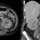
Unilateral striated nephrogram
- acute ureteric obstruction
- acute pyelonephritis
- acute renal vein thrombosis
- acutely following renal contusion
- acute radiation therapy to the kidney
Bilateral striated nephrograms
- autosomal recessive polycystic kidney disease (ARPCKD)
- acute pyelonephritis
- acute tubular obstruction
- acute tubular necrosis
- hypotension
- idiopathic asymptomatic in some children
History and etymology
The term "nephrogram" was originally used in the evaluation of plain film excretory urography . The use of intravenous contrast for imaging evaluation of the kidney was pioneered by Jewish-American physician Moses Swick (1900-1985) in 1929 . He worked with German urologist Alexander von Lichtenberg and chemist Arthur Binz to develop the first excretory contrast agent, known as Uroselectan.
In modern radiology, the nephrogram patterns are more commonly observed on CT urography and on plain film radiography following intravenous injection of an iodinated contrast agent.
Differential diagnosis
- spotted nephrogram
- more segmental or subsegmental areas of corticomedullary hypoattenuation
- secondary to multiple small-vessel infarctions
- seen in intrarenal vasculitis (classic), embolic disease
- see also: cortical rim sign
- rim nephrogram
- enhancing cortex, and absent medullary enhancement
- seen days-to-weeks following global renal infarction, due to collateral circulation
- main renal artery occlusion
- main renal vein occlusion/thrombosis (more commonly associated with enlarged kidney)
- see also: cortical rim sign
- reverse rim nephrogram
- absent cortical enhancement, and normal medullary enhancement
- indicative of acute cortical necrosis
Siehe auch:
- akute Pyelonephritis
- Pyelonephritis
- Nierenvenenthrombose
- autosomal recessive polycystic kidney disease (ARPKD)
- Nephritis
- acyclovir induced nephropathy
- Streifiges Nephrogramm als Zufallsbefund nach Kontrastmittelgabe bei Kindern in MRT
und weiter:


 Assoziationen und Differentialdiagnosen zu striated nephrogram: