renal cortical necrosis

Renal cortical necrosis occurs as a result of severe systemic illness in a variety of settings and can result in permanent renal impairment.
Pathology
Etiology
- severe hemodynamic shock
- traumatic blood loss
- postpartum hemorrhage
- septic shock
- venom toxin
- transfusion reaction
- severe dehydration
- microangiopathic hemolysis
- renal transplantation
Radiographic features
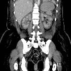
CT
Contrast-enhanced CT demonstrates a non-enhancing renal cortex and a normal enhancing renal medulla (reverse rim sign). A very thin rim of contrast enhancement (cortical rim sign) may persist and should not be mistaken for adequate perfusion.
Eventually dystrophic calcification of the renal cortex may be seen (cortical nephrocalcinosis).
MRI
Low signal intensity on both T1 and T2 weighted sequences affecting the inner renal cortex and the columns of Bertin is the major characteristic finding of renal cortical necrosis .
Additional features include:
- swelling of both kidneys
- corticomedullary differentiation seen better on T2-weighted images instead of T1-weighted images
Note that the cortical rim sign can persist in renal cortical necrosis because of its separate capsular blood supply.
Differential diagnosis
- Other causes of low signal intensity renal parenchyma on MRI
Siehe auch:
und weiter:


 Assoziationen und Differentialdiagnosen zu renal cortical necrosis: