perirenaler Abszess


A perinephric abscess may result due to rupture of a renal abscess into the perirenal space, but usually it develops directly from acute pyelonephritis. However, any inflammatory process outside the Gerota's fascia may also result in perinephric abscess. Perinephric abscesses are associated with diabetic patients with calculi and in patients with septic emboli.
Radiographic features
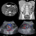
Ultrasound
Ultrasound is usually the first imaging investigation performed for assessment of a renal abscess, perinephric collection, or pyelonephritis. As elsewhere, the collection is usually hypoechoic or mixed echogenicity, depending on the content. Ultrasound may also aid treatment in offering percutaneous drainage of the collection.
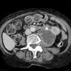
CT
Often shows areas of soft-tissue or fluid attenuation within the perirenal space. It may extend to involve psoas muscle and down to the pelvis. Gas may be present within the abscess.
Treatment and prognosis
Perinephric and mixed perinephric/renal abscesses are typically treated with imaged guided percutaneous drainage. This may be with either ultrasound-guided percutaneous abscess drainage or CT guided dependent on the visibility and accessibility. Only those with unsuccessful or inadequate treatment with percutaneous drainage and antibiotics tend to require surgery.
Siehe auch:
- perirenal fascia
- perirenal space
- akute Pyelonephritis
- Nierenabszess
- perirenales Hämatom
- perirenale Flüssigkeit
- Antibiom der Niere
- spontanes perirenales Hämatom
- calculi
- ultrasound-guided percutaneous abscess drainage
und weiter:


 Assoziationen und Differentialdiagnosen zu perirenaler Abszess: