emphysematöse Pyelonephritis


Emphysematous pyelonephritis (plural: emphysematous pyelonephritides) refers to a morbid infection with particular gas formation within or around the kidneys. If not treated early, it may lead to fulminant sepsis and, therefore, carries a high mortality.
Clinical presentation
The patient usually presents with fevers and flank pain. In diabetics, who are the ones at risk for this condition, leukocytosis and hyperglycemia are prominent laboratory findings.
The presence of thrombocytopenia is particularly associated with poor prognosis .
Pathology
Etiology
It tends to be more common in females, and approximately 90% of patients have uncontrolled diabetes mellitus . It may however also be seen in immunocompromised individuals or associated with urolithiasis, neoplasms, or sloughing of papilla.
Causative organisms include:
- Escherichia coli: usually considered the commonest causative organism
- Klebsiella pneumonia
- Proteus mirabilis
Radiographic features
Plain radiograph
May show mottled gas within the renal fossa or crescentic gas collection within Gerota's fascia. Linear gas shadows along paraspinal region may also be seen, representing retroperitoneal gas.
Ultrasound
- may show an enlarged kidney with coarse echoes within renal parenchyma or collecting system
- dirty echogenic foci with reverberation/ring-down artifacts representing gas ('dirty shadowing') may also be seen
CT
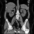
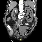
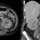
CT is the best diagnostic modality for emphysematous pyelonephritis, and it may show the following diagnostic features:
- enlarged, destroyed renal parenchyma
- small bubbly or linear streaks of gas
- fluid collections, with gas-fluid levels
- focal necrotic areas +/- abscess
Radiological classification
CT features of emphysematous pyelonephritis can be differentiated into two types :
- type 1
- greater than one-third renal parenchymal destruction
- streaky or mottled appearance of gas
- intra- or extrarenal fluid collections are characteristically absent
- it is usually more aggressive and lead to death shortly, if not intervened early
- mortality 70%
- type 2
- destruction of less than one-third of the parenchyma
- renal or extrarenal collections associated with bubbly or loculated gas, or gas within pelvicalyceal system or ureter
- mortality 20%
In addition to this, the Huang-Tseng CT classification system is also described:
- class 1: gas in the collecting system only
- class 2: gas in renal parenchyma only (without extrarenal extension)
- class 3: gas in renal parenchyma with extrarenal extension
- class 3a: extension of gas or abscess to perinephric space
- class 3b: extension of gas or abscess to pararenal space
- class 4: bilateral emphysematous pyelonephritis or solitary kidney with emphysematous pyelonephritis
Treatment and prognosis
In mild cases, treatment is with intravenous antibiotics. Percutaneous catheter drainage of perirenal or retroperitoneal collections can be performed. Severe cases may warrant nephrectomy.
Differential diagnosis
General imaging differential considerations include:
- emphysematous pyelitis
- iatrogenic (instrumentation, or intervention of urinary tract)
- fistulous communication with bowel
See also
Siehe auch:
und weiter:

 Assoziationen und Differentialdiagnosen zu emphysematöse Pyelonephritis:


