Pyramidal lobe of thyroid
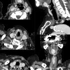


The pyramidal lobe of thyroid (a.k.a. Lalouette pyramid ) is a normal anatomic variant representing a superior sliver of thyroid tissue arising from the thyroid isthmus. It is seen as a third thyroid lobe and is present in 10-30% of the population.
It represents a persistent remnant of the thyroglossal duct. It usually arises from the right or left side of the isthmus extending in a cranial direction; pyramidal lobes arising directly from the midline of the isthmus were rare in a large ultrasound study, accounting for only 2% of the cases .
It is not uncommon to see it on routine thyroid ultrasound, a study of 416 patients in 2014 found it in 21% .
All the pathologies that may be seen in the normal thyroid are also seen in the pyramidal lobe.
A band of fibrous tissue may be present extending superiorly from the pyramidal lobe to the hyoid bone, sometimes with a skeletal muscle component, termed the levator glandulae thyroideae muscle .
Siehe auch:
und weiter:


 Assoziationen und Differentialdiagnosen zu Lobus pyramidalis: