thyroid gland


The thyroid gland is a single midline endocrine organ in the anterior neck responsible for thyroid hormone production which lies in the visceral space completely enveloped by pretracheal fascia (middle-layer of the deep cervical fascia).
Gross anatomy
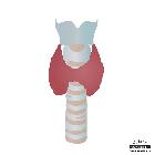
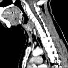
The thyroid extends from C5 to T1 and lies anterior to the thyroid and cricoid cartilages of the larynx and the first five or six tracheal rings.
The thyroid is butterfly or "H"-shaped and is composed of two lobes, each with a superior and inferior pole. Usually, the superior pole is narrower than the inferior pole giving a pear-like shape to each lateral lobe. The lateral lobes are connected in the midline by a narrow isthmus which is adherent to the 2nd-4th tracheal rings. Each lobe measures approximately 4 cm in length. Average weight is 25 g; this is slightly higher in females and may increase during menstruation and pregnancy .
The parathyroid glands lie posteromedially and are sometimes intracapsular.
The ligament of Berry is a posterior extension of the thyroid capsule which attaches to the cricoid cartilage and the upper tracheal rings. It encloses a short segment of the recurrent laryngeal nerve as it ascends in the tracheo-esophageal groove. As such it is an important surgical landmark during thyroidectomies to avoid damaging the nerve.
Relations
- anteriorly: strap muscles
- posteriorly: thyroid cartilage, cricoid cartilage, trachea
- posteromedially: tracheo-esophageal groove (containing lymph nodes, recurrent laryngeal nerve, parathyroid glands)
- posterolaterally: carotid space
Arterial supply
- superior thyroid artery (from the external carotid artery)
- inferior thyroid artery (from the thyrocervical trunk)
- if the inferior thyroid artery arises from the subclavian artery it is referred to as an accessory inferior thyroid artery
Venous drainage
- superior thyroid vein (drains to the internal jugular vein)
- middle thyroid vein (drains to the internal jugular vein)
- inferior thyroid vein (drains via plexus to the brachiocephalic vein)
Lymphatic drainage
Lymphatic drainage is multidirectional and initial lymph drainage is to perithyroid lymph nodes then onto prelaryngeal, pretracheal and paratracheal nodes (level 6 lymph nodes).
Innervation
Sympathetic supply is provided by superior, middle and inferior cervical ganglia.
Embryology
The thyroid gland develops from the proximal primitive foregut between the first and second pharyngeal pouches at the foramen cecum, in the midline of the base of the tongue. During the 5th embryonic week, a diverticulum forms at the foramen which inferiorly migrates anterior to the body of the hyoid bone, curving posterior and superiorly to reach behind the bone before once more turning inferiorly and continuing anterior to the larynx, forming the thyroglossal duct . The tip of the duct bifurcates, forming the two lobes of the gland. The parafollicular cells (C cells) responsible for calcitonin production are derived from separate tissue, the ultimobranchial body, a small diverticulum of the fourth pharyngeal pouch .
Variant anatomy
- lobar hemiagenesis
- pyramidal lobe
- superiorly-projecting thyroid tissue from the isthmus
- thyroglossal duct cyst
- ectopic thyroid tissue
- accessory thyroid gland
- Zuckerkandl's tubercle
- the gland may be supplied by a thyroidea ima artery, which may replace the inferior thyroid artery (3%)
Radiographic appearance
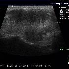
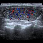
Ultrasound
- the normal thyroid gland has a homogeneous appearance
- the capsule may appear as a thin hyperechoic line
- each lobe normally measures
- length: 4-7 cm
- depth: <2 cm
- isthmus <0.5 cm deep
- volume (excluding isthmus, unless its thickness is >3 mm)
- 10-15 mL for females
- 12-18 mL for males
Related pathology
Neoplasms
Inflammatory conditions: thyroiditis
Autoimmune
- Graves disease
- de Quervain thyroiditis (subacute granulomatous)
- postviral
- associated with HLA-B35
- acutely there is an increase in thyroid hormone, with a resultant decrease in TSH. As a result there is a decrease in I uptake; eventually, however, the main phase is that of a hypothyroid state
- subacute lymphocytic thyroiditis
- painless
- young women, especially in postpartum period
- gland is usually normal in size, or minimally increased
- usually an early hyperthyroid state which returns to normal, but may have a transient late hypothyroid period
- Hashimoto thyroiditis
- F>M 10:1
- associated with
- Down syndrome and Turner syndrome
- autoimmune conditions including systemic lupus erythematosus, Sjögren syndrome and myasthenia gravis
- primary thyroid lymphoma
- anti-TSH-receptor-autoantibodies
- CD8+ cytotoxic T-cell mediated
- usually hypothyroid, although there may be a brief hyperthyroid early phase
- Riedel thyroiditis
- distinctive in that inflammation extends beyond the confines of the gland in to adjacent tissues
- typically presents as a hard goiter which commonly compresses the trachea; it is hypoenhancing and hypoechogenic
- associated with:
Note: although Graves disease is autoimmune it is not really a thyroiditis as it does not have a significant inflammatory component.
Infective
Includes thyroiditis associated with:
- PCP/PJP
- MAIC
- fungal
Others
History and etymology
"Thyroid" derives from the Greek word "θνρεός" (large oblong shield) and "είδος" (-like) . The Greek shield had a notch cut into it for the chin, and the resemblance of the shield to a particular piece of cartilage in the neck brought about the term "thyreoid cartilage" (the "e" was later dropped).
The thyroid gland was originally known as the "laryngeal gland", and was renamed the "thyroid gland" by Thomas Wharton in 1646.
Siehe auch:
- Morbus Basedow
- Primär sklerosierende Cholangitis
- Hashimoto-Thyreoiditis
- Subakute Thyreoiditis de Quervain
- Zungengrundstruma
- Neoplasien der Schilddrüse
- Thyreoiditis
- thyroid cancer staging
- Riedel-Thyreoiditis
- retroperitoneale Fibrose allgemein
und weiter:
- laterale Halszyste
- Thymus
- Arteria carotis communis
- Tumoren des vorderen oberen Mediastinums
- Schilddrüsenknoten
- Struma
- ektopes Schilddrüsengewebe
- Ductus thyreoglossus
- thyroid scan (Tc-99m)
- Arteria thyroidea ima
- follikuläres Schilddrüsenkarzinom
- Zyste oder Fistel des vierten Kiemenbogens
- retrosternale Struma
- Zyste oder Fistel des ersten Kiemenbogens
- thyroidales Inferno
- zervikale Mittellinienraumforderungen
- Arteria thyroidea superior
- Vena thyroidea inferior


 Assoziationen und Differentialdiagnosen zu Schilddrüse: