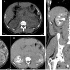
renal involvement with lymphoma

Spectrum of
imaging findings in AIDS-related diffuse large B cell lymphoma. Ultrasound image of the left kidney in a HIV-positive male demonstrates heterogeneously echogenic areas in the medulla extending to the collecting system. Unenhanced CT KUB showed diffuse thickening of the renal pelvis with hyperdense soft tissue material relative to the renal parenchyma. Heterogenous infiltration of the kidney is seen on the postcontrast CT images in arterial and urographic phases

Diffuse large
B-cell lymphoma with bilateral renal involvement. Axial T2-weighted MRI demonstrating rounded areas of hypointensity involving the lateral right upper pole and medial left upper pole of the right and left kidneys, respectively (arrows).

Diffuse large
B-cell lymphoma with bilateral renal involvement. Rotating MIP image demonstrating intensely increased uptake in the liver mass (arrowhead), upper pole of the right kidney (+), and throughout the left renal parenchyma (arrows).

Diffuse large
B-cell lymphoma with bilateral renal involvement. Fused Coronal PET-CT demonstrating intensely increased uptake in the lateral upper pole of the right kidney (arrow).

