retroaortaler Verlauf der linken Nierenvene

Retroaortic
left renal vein • Retroaortic left renal vein - Ganzer Fall bei Radiopaedia

Retroaortic
left renal vein • Retroaortic left renal vein - Ganzer Fall bei Radiopaedia

Retroaortaler
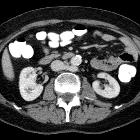
Verlauf der linken Nierenvenen in der Computertomographie. Häufige Variante.

Retroaortic
left renal vein • Calyceal diverticulum - Ganzer Fall bei Radiopaedia

Anatomical
variant of a duplicated retroaortic left renal vein: Clinical manifestation, radiographic features and management. Axial view of enhanced abdominal CT with IV contrast demonstrating the inferior branch (green arrow) of the duplicated retroaortic left renal vein, with the renal vein visualized between the aorta and lumbar vertebrae.

Anatomical
variant of a duplicated retroaortic left renal vein: Clinical manifestation, radiographic features and management. Axial view of enhanced abdominal CT with IV contrast demonstrating the superior branch (red arrow) of the duplicated retroaortic left renal vein, with the renal vein visualized between the aorta and lumbar vertebrae.

Retroaortic
left renal vein • Traumatic renal injury - AAST grade IV injury - Ganzer Fall bei Radiopaedia

Retroaortic
left renal vein • Retroaortic left renal vein - Ganzer Fall bei Radiopaedia

Azygos
continuation of the inferior vena cava • Azygous continuation of the inferior vena cava - Ganzer Fall bei Radiopaedia

Retroaortic
left renal vein • Retroaortic left renal vein: type III - Ganzer Fall bei Radiopaedia

Retroaortic
left renal vein • Retroaortic left renal vein - Ganzer Fall bei Radiopaedia

Retroaortic
left renal vein • Circumaortic left renal vein - Ganzer Fall bei Radiopaedia

Retroaortic
left renal vein • Duplicated left renal vein - Ganzer Fall bei Radiopaedia

Retroaortic
left renal vein • Retroaortic left renal vein - Ganzer Fall bei Radiopaedia

Inferior vena
cava • Caval variants (illustrations) - Ganzer Fall bei Radiopaedia

Anatomical
variant of a duplicated retroaortic left renal vein: Clinical manifestation, radiographic features and management. Coronal view of enhanced abdominal CT with IV contrast demonstrating superior (red arrow) and inferior (green arrow) branches of the duplicated retroaortic left renal vein.

Retroaortic
left renal vein • Type IV left renal vein - Ganzer Fall bei Radiopaedia

Retroaortic
left renal vein • Retroaortic left renal vein - type II - Ganzer Fall bei Radiopaedia

Retroaortic
left renal vein • Retroaortic left renal vein - Ganzer Fall bei Radiopaedia

Retroaortic
left renal vein • Retroaortic left renal vein (MRI) - Ganzer Fall bei Radiopaedia

Teilweise
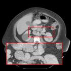
retroaortaler Verlauf der linken Nierenvenen: oberes Bild schmaler präaortaler Anteil, unteres Bild mit retroaortalem Anteil. Computertomographie axial.

Hemoperitoneum
• Ruptured corpus luteum cyst with hemoperitoneum - Ganzer Fall bei Radiopaedia
Retroaortic left renal vein (RLRV) is a normal anatomical variant where the left renal vein is located between the aorta and the vertebra and drains into the inferior vena cava.
Its recognition is important in order to avoid complications during retroperitoneal surgery or interventional procedures .
Epidemiology
Retroaortic left renal vein has an estimated prevalence of ~2% .
Clinical presentation
Urological symptoms can be caused by increased pressure in the renal vein resulting in venous hypertension. This is an atypical form of nutcracker syndrome. Patients can present with hematuria and recurrent left flank pain.
Types
The retroaortic position of the left renal vein has four subtypes :
Siehe auch:
und weiter:

 Assoziationen und Differentialdiagnosen zu retroaortaler Verlauf der linken Nierenvene: