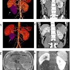
retroperitoneales Hamartom

Imaging
findings for pancreatic Hamartoma: two case reports and a review of the literature. The lesion showed a hypoechoic mass in the pancreatic head on US (a). Axial CT of the abdomen (plain scan and venous phase) showing a 2.9-cm iso-density lesion in the pancreatic head that was well-defined after enhancement (b-c). Axial pancreatic MRI showing a solid lesion with several small cystic lesions, T1WI showed hypointensity (d), T2WI showed iso- to high-intensity (e), and DWI showed iso-intensity (F); the lesion had obvious progressive enhancement (g-i)

Retroperitoneal
hamartoma mimicking angiomyolipoma: a case report and review of literature. A–D A large soft tissue dense mass (white arrow) was seen below the right kidney of the retroperitoneum on CT images, with an ill-defined boundary with the psoas major muscle. No definite enhancement was observed after iodine contrast injection. E MRI T1WI showed hyperintense signal mass with ill-defined boundary with psoas major muscle and flowing void effect was revealed in the tumor. F After gadolinium injection, cluster and speckled enhanced focal shadows were observed in the mass on coronal MRI

Imaging
findings for pancreatic Hamartoma: two case reports and a review of the literature. The lesion showed a hypoechoic mass in the pancreatic head on US (a). Axial CT of the abdomen (plain scan and venous phase) showing a 1.4-cm iso-density lesion in the pancreatic head that was well-defined after enhancement (b-c). Axial pancreatic MRI showing a solid lesion, T1WI showed hypointensity (d), T2WI showed iso-intensity (e), DWI showed iso-intensity (f), the lesion had obvious progressive enhancement (g-i)
retroperitoneales Hamartom
Siehe auch:

 Assoziationen und Differentialdiagnosen zu retroperitoneales Hamartom:


