Hamartom des Pankreas

Imaging
findings for pancreatic Hamartoma: two case reports and a review of the literature. The lesion showed a hypoechoic mass in the pancreatic head on US (a). Axial CT of the abdomen (plain scan and venous phase) showing a 2.9-cm iso-density lesion in the pancreatic head that was well-defined after enhancement (b-c). Axial pancreatic MRI showing a solid lesion with several small cystic lesions, T1WI showed hypointensity (d), T2WI showed iso- to high-intensity (e), and DWI showed iso-intensity (F); the lesion had obvious progressive enhancement (g-i)

Imaging
findings for pancreatic Hamartoma: two case reports and a review of the literature. The lesion showed a hypoechoic mass in the pancreatic head on US (a). Axial CT of the abdomen (plain scan and venous phase) showing a 1.4-cm iso-density lesion in the pancreatic head that was well-defined after enhancement (b-c). Axial pancreatic MRI showing a solid lesion, T1WI showed hypointensity (d), T2WI showed iso-intensity (e), DWI showed iso-intensity (f), the lesion had obvious progressive enhancement (g-i)

A typical
case of resected pancreatic hamartoma: a case report and literature review on imaging and pathology. Dynamic computed tomography (CT) showed hypodensity in the arterial phase (1a), slight enhancement in the portal phase and iso- to hyper-density in the equilibrium phase (1b), compared to the parenchyma of the pancreas (1c), each indicated with arrowheads

A typical
case of resected pancreatic hamartoma: a case report and literature review on imaging and pathology. Magnetic resonance imaging (MRI) showed high signal intensity in T2WI (2a) and low intensity in T1WI (2b), each indicated with arrowheads

A typical
case of resected pancreatic hamartoma: a case report and literature review on imaging and pathology. Endoscopic ultrasonography (EUS) depicted well-demarcated mass (indicated with arched line) with inside mosaic pattern (3a), and the mass was described harder than the parenchyma of the pancreas by elastography (3b)

Pancreatic
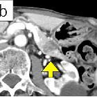
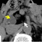
hamartoma: a case report and literature review. Chronological changes seen on CT. a: First examination. The hypo-enhanced mass was 4.2 × 3.9 cm in size with solid and cystic lesions located in the uncus of the pancreas. b: At 21 months after first examination. The tumor shown is 3.9 × 3.6 cm in size. The cystic lesion (yellow arrow) had become smaller and the solid lesion (white arrow) had become larger. c: At 28 months after first examination. The tumor was 4.2 × 3.3 cm in size. The cystic lesion changed and displayed irregular margins

Pancreatic
hamartoma: a case report and literature review. a, b, c. Chronological changes seen on MRCP. a: At the first examination. The tumor was observed as multiple cystic lesions. b: At 21 months after first examination. c: At 28 months after first examination. Figure 2-d, e, f, g. MRI appearance at 28 months after first examination. d: T1WI. A multi-cystic lesion with low intensity was surrounded by a mass with iso-intensity. e: T2WI. A multi-cystic lesion displays low- to iso-intensity. The surrounding tissue shows iso- to high intensity. f: T2-FAT-SAT. A multi-cystic high intensity lesion is shown. The surrounding tissue shows complete fat suppression. g: T2WI (coronal image). A ringed multi-cystic lesion with high intensity is surrounded by a smooth superficial mass. A small nodule is seen inside of the cyst ring

Pancreatic
hamartoma: a case report and literature review. Intraoperative ultrasound examination. A well-demarcated honeycomb-like cystic lesion in the pancreatic uncus was found. There was no evidence of venous or ductal invasion
 Assoziationen und Differentialdiagnosen zu Hamartom des Pankreas:


