benigne Pankreastumoren

Imaging
findings for pancreatic Hamartoma: two case reports and a review of the literature. The lesion showed a hypoechoic mass in the pancreatic head on US (a). Axial CT of the abdomen (plain scan and venous phase) showing a 2.9-cm iso-density lesion in the pancreatic head that was well-defined after enhancement (b-c). Axial pancreatic MRI showing a solid lesion with several small cystic lesions, T1WI showed hypointensity (d), T2WI showed iso- to high-intensity (e), and DWI showed iso-intensity (F); the lesion had obvious progressive enhancement (g-i)

Zufallsbefund
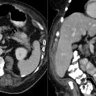
in der Computertomographie: Lipom des Pankreas. Umschriebener fett-isodenser Herd im Parenchym des Pankreas.

Lipom des
Pankreas (im Gegensatz zur Pankreaslipomatose). Seltener Zufallsbefund hier in der Computertomographie axial und coronar. (Zusätzlich Verdacht auf Empyem der Pleura rechts.)

Imaging
findings for pancreatic Hamartoma: two case reports and a review of the literature. The lesion showed a hypoechoic mass in the pancreatic head on US (a). Axial CT of the abdomen (plain scan and venous phase) showing a 1.4-cm iso-density lesion in the pancreatic head that was well-defined after enhancement (b-c). Axial pancreatic MRI showing a solid lesion, T1WI showed hypointensity (d), T2WI showed iso-intensity (e), DWI showed iso-intensity (f), the lesion had obvious progressive enhancement (g-i)
benigne Pankreastumoren
Siehe auch:
und weiter:

 Assoziationen und Differentialdiagnosen zu benigne Pankreastumoren:


