Rheumatoid nodules

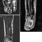
Pseudotumoural
soft tissue lesions of the foot and ankle: a pictorial review. Rheumatoid nodule. Short-axis T1-WI (a), fat-suppressed T2-WI (b): nodular lesion at the plantar subcutaneous fat tissue at the level of the metatarsals. The lesion is of low SI on T1-WI, whereas heterogeneity is seen on T2-WI. Multiple erosions were seen at the metatarsophalangeal joints (not shown)
Pseudotumoural
soft tissue lesions of the hand and wrist: a pictorial review. Histopathologically proven rheumatoid nodule in a patient with known rheumatoid arthritis. a Coronal SE T1-WI. Hypointense subcutaneous nodule at the palmar aspect of the distal phalanx of the right digit 3 (arrowheads). b Sagittal FS TSE T2-WI. High signal intensity of the lesion (black arrows). There is moderate pressure erosion of the palmar aspect of the distal phalanx. c Sagittal FS SE T1-WI after intravenous injection of gadolinium contrast medium. There is peripheral enhancement of the lesion (black arrowheads). Palmar localisation of a rheumatoid nodule is less frequent than localisation at the dorsal aspect of the hand
The term rheumatoid nodules can either mean
Siehe auch:


 Assoziationen und Differentialdiagnosen zu