Schlüsselbein


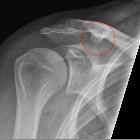
The clavicle, also colloquially known as the collarbone, is the only bone connecting the pectoral girdle to the axial skeleton and is the only long bone that lies horizontally in the human skeleton.
Gross anatomy
Osteology
The clavicle is roughly "S-shaped" with a flattened, concave, lateral one-third and a thickened, convex, medial two-thirds. On the inferior surface of lateral third is the conoid tubercle for the attachment of the conoid ligament and lateral to this is the trapezoid line for attachment of the trapezoid ligament. On the inferior surface of the medial clavicle is the costal tuberosity and groove for subclavius for the attachment of costoclavicular ligament and subclavius muscle, respectively.
The female clavicle is shorter, thinner and less curved than the male clavicle.
Articulations
The clavicle articulates with the acromion at the acromioclavicular joint laterally and the sternum at the sternoclavicular joint medially.
Attachments
- muscles
- pectoralis major, sternocleidomastoid (clavicular head), deltoid, trapezius, subclavius, sternohyoid
- ligamentous
- acromioclavicular ligament, coracoclavicular ligament, sternoclavicular ligament, costoclavicular ligament, interclavicular ligament
Arterial supply
- nutrient branch from the suprascapular artery
Development
Ossification
It is the first bone to start ossification at around 5th-6th weeks of gestation. It is also the last ossification center to fuse, around 22-25 years of age. The lateral end has intramembranous ossification. See main article: ossification centers of the pectoral girdle.
Variant anatomy
- forked clavicle
- supraclavicular foramen: the clavicle may be pierced by a branch of supraclavicular nerve
- coracoclavicular joint
- hypertrophic conoid tubercles
- At the attachment of the costoclavicular (rhomboid) ligament, there may be a tuberosity or depression (rhomboid fossa) of variable size that may mimic disease. A rhomboid fossa is more common in younger adults and males . See case 5.
Radiographic features
Plain radiograph
On a chest x-ray image, the clavicles are superimposed over the apex of both the lungs and obscure the subtle lesions. An apical or lordotic view may then provide greater detail of the lung apices.
Chest x-rays are correctly aligned if the medial ends of clavicles are equidistant from the spinous process of vertebrae at the T4/5 level.
Related pathology
- clavicular fracture: the weakest part of clavicle is the junction of lateral one third and medial two third and this is the most common site of fracture
- acromioclavicular joint injury
- sternoclavicular joint dislocation
- clavicle tumors
- sclerotic clavicle
- pediatric clavicle abnormalities
- osteolysis
- distal clavicular erosions
Siehe auch:
- Tuberculum conoideum der Klavikula
- Klavikulafraktur
- clavicle abnormalities (paediatric)
- AC-Gelenksprengung
- Musculus subclavius
- Geburtstraumatische Claviculafraktur
- acromioclavicular arthrosis
- Tumoren der Klavikula
- normal clavicle radiograph
- absent clavicles
- Klavikulektomie
und weiter:

 Assoziationen und Differentialdiagnosen zu Klavikula:


