Schockdarm

Infant who
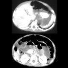
became unresponsive and underwent 30 minutes of CPRAxial CT with contrast of the abdomen shows periportal edema in the liver, diffusely dilated and hyperenhancing loops of small bowel and a narrow caliber inferior vena cava.The diagnosis was hypoperfusion complex in a child abuse patient.

Intrathoracic
viscera herniation and other thoracoabdominal injuries in a young female multitrauma patient: Computed Tomography evaluation. Intraperitoneal free fluid, thickened small bowel loops (on the left) and collapsed I.V.C. (shock bowel findings).
Shock bowel is the appearance of the bowel in a state of hypotension. It is usually seen as part of the CT hypoperfusion complex.
Radiographic features
CT
- thickened bowel loops (>3 mm) with enhancing walls (the reason the condition was previously known as "shock bowel")
- on non-contrasted images, hyperdense walls compared to the psoas muscle
- wall thickening is due to submucosal edema
- hyperenhancing mucosa
Siehe auch:
- Mesenterialinfarkt
- Chronisch-entzündliche Darmerkrankungen
- CT Hypotensionskomplex
- radiation enteritis
- IgA-Vaskulitis
und weiter:

 Assoziationen und Differentialdiagnosen zu Schockdarm:
Chronisch-entzündliche
Darmerkrankungen


