CT Hypotensionskomplex


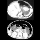
CT hypoperfusion complex refers to the predominantly abdominal imaging features that occur in the context of profound hypotension. Multiple abdominal organs can display atypical appearances not related to the initial trauma but reflect alterations in perfusion secondary to hypovolemia which affects the sympathetic splanchnic stimulation. The small bowel is more commonly affected than the large bowel.
Terminology
The term CT hypoperfusion complex is now preferred over the older and less accurate term shock bowel.
Pathology
Etiology
CT hypoperfusion complex is most commonly described in the context of post-traumatic hypovolemic shock but can also occur in :
- sepsis
- severe head or spinal injury
- cardiac arrest
- bacterial endocarditis
- diabetic ketoacidosis
Radiographic features
CT
Features of CT hypoperfusion complex have been defined as (some or all of these features may be present):
- small caliber abdominal aorta: AP diameter <13 mm both 20 mm above and below the renal arteries ; this occurs in ~30% of patients with hypovolemia, and is not specific
- collapsed inferior vena cava: AP diameter <9 mm in three consecutive segments; i.e. 20 mm both above and below the renal veins and the perihepatic portion
- sometimes not appreciated due to massive fluid resuscitation
- halo sign
- low density (<20 HU) fluid surrounding the IVC
- this occurs in ~80% of patients with severe hypovolemia
- thickened bowel loops (>3 mm) with enhancing walls (the reason the condition was previously known as "shock bowel")
- most commonly involves the jejunum
- on non-contrasted images, hyperdense walls compared to the psoas muscle
- wall thickening is due to submucosal edema
- hyperenhancing mucosa
- shock pancreas: heterogeneous enhancement with peripancreatic fluid (<20 HU) ; this is a controversial finding
- hypoenhancement of the spleen (subjective)
- decreased splenic volume
- hypoenhancement of the liver: 25 HU less than the spleen
- bilateral adrenal gland hyperenhancement may be a feature mainly in pediatrics and is controversial in adults
- hyperenhancing kidneys
- ascites
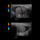
- shock thyroid: heterogenous contrast enhancement with enlargement of the thyroid gland which may mimic a multinodular gland
The small bowel findings are the most commonly observed feature in CT hypoperfusion complex. Note that the frequency of most associated findings does not differ between trauma and non-trauma patients with CT hypoperfusion complex, except in case of a collapsed IVC which occurs in the majority of trauma patients, but in <40% of non-trauma patients with CT hypoperfusion complex . A follow-up study following resuscitation may demonstrate reversal of small bowel findings.
In the neck, an uncommon finding is shock thyroid, where the thyroid gland demonstrates heterogeneous thyroid contrast enhancement and perithyroid fluid .
Treatment and prognosis
CT findings tend to be reversible with appropriate fluid management although severe hypotension and shock have a significant mortality rate.
Differential diagnosis
The clinical context is extremely valuable for image interpretation. The differential for thickened enhancing bowel includes :
- idiopathic angioedema
- hereditary angioedema
- ischemic bowel
- vasculitides (e.g. Henoch-Schonlein purpura)
- infective enteritis
- inflammatory bowel disease
- radiation enteritis
- submucosal or intramural hemorrhage (e.g. coagulopathy)
Siehe auch:


 Assoziationen und Differentialdiagnosen zu CT Hypotensionskomplex: