Second branchial cleft fistula


Second branchial cleft fistulae are congenital anomalies of embryonic development of branchial apparatus with the external cutaneous ostium in the lateral neck connecting to the tonsillar fossa. They can be diagnosed as a result of typical clinical presentation and the diagnosis can be confirmed various imaging modalities, including fistulography, ultrasound, CT and MRI.
Epidemiology
Second branchial cleft fistulae are rare and comprise only 2% of all branchial anomalies. They are almost always present at birth, however the small pinpoint external opening may go unnoticed. These individuals, commonly present in the first and second decade of life. There is a slight with male predilection (M:F 3 : 2)
Only 39% are complete fistulae, linking the skin to the pharynx, with the majority (50%) only having a draining sinus; 11% have internal opening alone .Bilateral fistulae found in 2-10% of cases. In patients with unilateral fistulae, 70% occurs on the right side .
Clinical presentation
Second branchial cleft fistulae typically open onto the skin at the anterior border of sternocleidomastoid at the junction of middle and lower 1/3.
These fistulae typically present with intermittent or continuous mucous discharges from the cutaneous opening at the lateral neck and may suffer from recurrent attacks of inflammation, particularly after a preceding upper respiratory tract infection . On occasion cellulitis or even abscess formation occur. Uncommonly (~10%) the lining of the fistula can undergo malignant degeneration into squamous cell carcinoma .
Due to the proximity of the track to the vagus nerve, instrumentation may result in symptoms such as cough, palpitation, pallor and vomiting .
Pathology
The embryology of the branchial apparatus is complicated and results in a variety of malformations described further in branchial cleft anomalies.
Second branchial cleft fistulous tracts are lined by keratinized squamous epithelium .
Radiographic features
Second branchial cleft fistulae pass deep to second arch structures and superficial to the third arch structures. As such the tract passes from the tonsillar fossae and passes between the internal and external carotid artery before opening onto the skin .
Fistulography
Fistulography (opacifying the fistula with contrast media) delineates a smooth tract extending from the external cutaneous opening at the lateral neck superomedially passing between the ECA and ICA to the tonsillar fossa.
Ultrasound
Ultrasound can demonstrate the fistulous tract as an anechoic linear structure filled with faint echogenic debris, comprising cellular material, cholesterol crystals and keratin.
CT
CT, particularly combined with fistulography offers excellent anatomical delineation.
MRI
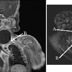
Less diagnostic value but T1 weight images provides better soft tissue contrast of the upper half of the fistula at the level of carotid bifurcation .
Treatment and prognosis
Due to the risk of recurrent infection and even squamous cell carcinoma obliteration of the tract is preferred. Options include: use of sclerosing agents; excision of the fistulous tract (Hockey stick type or Step Ladder incision) andpull through brachial fistulectomy.
Siehe auch:
- Schwannom
- zystische Lymphknoten
- Neurofibrom
- Ductus thyreoglossus Zyste
- lymphoepitheliale Zyste der Glandula parotis
- Zyste oder Fistel des zweiten Kiemenbogens
- zervikale bronchogene Zyste
- benignes Ganglioneurom
- tuberkulöse Halslymphknoten
- Halsfistel
- Kiemenbogenzyste
- Zyste oder Fistel des vierten Kiemenbogens
- Kiemenbogen


 Assoziationen und Differentialdiagnosen zu Fistel des zweiten Kiemenbogens: