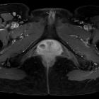
skene duct cyst
Paraurethral duct cysts are retention cysts that form secondary to inflammatory obstruction of the paraurethral (Skene) ducts in females.
Pathology
The cysts are lined by stratified squamous epithelium due to their origin from the urogenital sinus.
Clinical presentation
Usually asymptomatic.
Radiographic features
MRI
Typically appear as round or oval masses that are located just laterally to the external urethral meatus and inferior to the pubic symphysis.
On MR imaging, Skene duct cysts manifest as round or oval hyperintense lesions just lateral to the external urethral meatus.
Signal characteristics include:
- T2
- hyperintense signal if uncomplicated
- may show fluid-fluid levels if there is a complicating hemorrhage
Treatment and prognosis
Large, symptomatic cysts may warrant surgical excision or marsupialisation, usually with excellent outcome. Large cysts may also require drainage or excision in the presence of superimposed infection.
Differential diagnosis
General considerations include:
- Bartholin gland cyst
- urethral diverticulum (urethrocele)
- tend to be midurethral in location versus paraurethral gland cysts, which are located near the external urethral meatus
See also
Siehe auch:
und weiter:

 Assoziationen und Differentialdiagnosen zu skene duct cyst:

