Bartholin-Zyste


Bartholin gland cysts (sometimes shortened to Bartholin cysts) are cysts of the Bartholin gland, found in the posterolateral inferior third of the vagina and are associated with the labia majora.
Clinical presentation
Most patients are asymptomatic .
Complications
- infection: may turn into Bartholin gland abscesses
- rare instances of development of adenocarcinoma or squamous cell carcinoma within the cyst
Pathology
Cysts form as a result of an obstruction of the gland's duct by a stone/stenosis related to prior infection or trauma . Chronic inflammation can lead to ductal obstruction from pus or thick mucus which in turn can result in retained secretions within the Bartholin glands.
Radiographic features
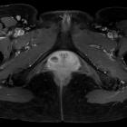
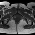
They are typically seen as rounded unilocular cysts lying at the posterior aspect of the vagina. Their location is at or below the level of the pubic symphysis (best appreciated on coronal imaging).
Ultrasound
May only be demonstrated on transperineal ultrasound if the cyst is close to the labia.
MRI
Signal characteristics include:
- T1: can be of variable signal
- T2: often of uniform hyperintensity on T2-weighted imaging although can occasionally vary dependent on protein content, may also be heterogeneous when infected
Treatment and prognosis
Initially, Bartholin cysts can be treated conservatively with pain management, warm soaks and warm compressions. If infected, oral antibiotics can be used.
If the infected cyst did not respond to antibiotics, surgical drainage can be considered in the form of incision and drainage or balloon catheter insertion.
The balloon catheter is inserted into the affected labia minora through the Bartholin cyst, and is left to drain into the vagina. The balloon catheter is left for four weeks to allow for a permanent epithelial connection between the Bartholin gland and the vagina .
Recurrent Bartholin's cysts may require marsupialization.
History and etymology
The Bartholin glands are named after the Danish anatomist Caspar Bartholin the Younger (1655-1738), who made the first detailed study of their physiology and anatomy in humans .
Differential diagnosis
General imaging differential considerations include:
- Bartholin gland abscess: may show associated inflammatory features
- Bartholin gland tumor: consider if discovered in the postmenopausal patient
- Gartner duct cyst: their location at or above the level of the pubic symphysis helps to differentiate them from Bartholin duct cysts
- Nabothian cyst: located at a much higher position within the uterine cervix
- Skene duct cyst: centered more anteriorly and closer to the external urethral meatus, sagittal imaging may help differentiate in some cases
- urethral diverticulum
Siehe auch:
- gartner duct cyst
- Ovula Nabothi
- Divertikel der Urethra
- skene duct cyst
- Abszess der Bartholinschen Drüsen
- Tumoren der Bartholinschen Drüsen
und weiter:

 Assoziationen und Differentialdiagnosen zu Bartholin-Zyste:

