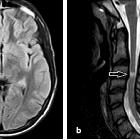
spinaler Befall bei Encephalomyelitis disseminata

Radiological
approach to non-compressive myelopathies. Multiple sclerosis in a 13-year-old female who presented with paraparesis. T2W axial MR image of the upper cervical spine (A) shows a peripherally located wedge shaped hyperintense lesion (white arrow) which involves less than half of the cross-sectional area of the spinal cord. Sagittal T2W image of the brain (B) of the same patient demonstrates patchy areas of T2 hyperintensity in pericallosal and periventricular distribution (black arrow). Sagittal T2W MR image of the cervical and upper thoracic spine (C) in another case of diagnosed MS shows a short segment hyperintense area within the cervical spinal cord (white arrows)

Location,
length, and enhancement: systematic approach to differentiating intramedullary spinal cord lesions. Multiple sclerosis. A 50-year-old male with a history of multiple sclerosis. a Axial MERGE image demonstrates partial cord involvement—focal hyperintensity within the right lateral cord with a characteristic triangular shape (arrow). There is no cord enlargement. b Sagittal T2 fat-saturated image demonstrates several scattered, short-segment foci of T2 hyperintensity (brackets)

Location,
length, and enhancement: systematic approach to differentiating intramedullary spinal cord lesions. Multiple sclerosis. A 50-year-old female with prior history of visual changes, presenting with right extremity weakness. a Sagittal STIR image shows several foci of short segment hyperintensity (brackets). b Axial T2 image demonstrates partial cord involvement, with a triangular lesion involving the right lateral cord (arrow). c Axial T1 post-contrast image demonstrates associated enhancement (arrow), consistent with active demyelination
spinaler Befall bei Encephalomyelitis disseminata
Siehe auch:
und weiter:

 Assoziationen und Differentialdiagnosen zu spinaler Befall bei Encephalomyelitis disseminata:

