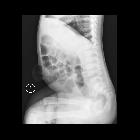
spontane Aszitesentleerung über Nabelhernie

Spontaneous
leakage of ascites from umbilical hernia in alcohol-related cirrhosis. Precontrast scans (a,b) showed cirrhotic liver, abundant homogeneously fluid-attenuating ascites (*), and umbilical hernia (arrow in b) contaning ascitic fluid.

Spontaneous
leakage of ascites from umbilical hernia in alcohol-related cirrhosis. Precontrast scans (a,b) showed cirrhotic liver, abundant homogeneously fluid-attenuating ascites (*), and umbilical hernia (arrow in b) contaning ascitic fluid.

Spontaneous
leakage of ascites from umbilical hernia in alcohol-related cirrhosis. Multiphase contrast-enhanced study including arterial (c,d), portal venous (e..h) and equilibrium (not shown) phases. No focal liver lesions suspicious for hepatocellular carcinoma were present. Note abundant ascites (*).

Spontaneous
leakage of ascites from umbilical hernia in alcohol-related cirrhosis. Multiphase contrast-enhanced study including arterial (c,d), portal venous (e..h) and equilibrium (not shown) phases. No suspicious focal liver lesions were present. Additionally, a right-sided inguinal hernia (arrowhead) filled with ascitic fluid (*) was present.

Spontaneous
leakage of ascites from umbilical hernia in alcohol-related cirrhosis. Portal venous phase images (e..h) confirmed abundant multicompartmental ascites (*), patent porto-mesenteric venous system, absence of suspicious focal liver lesions, and a right-sided inguinal hernia (arrowhead) filled with ascitic fluid.

Spontaneous
leakage of ascites from umbilical hernia in alcohol-related cirrhosis. Detail multiplanar contrast-enhanced images (f...h) showed round-shaped, externally convex umbilical hernia (arrows) filled with ascitic fluid, with narrow neck and integrity of the uniform, enhancing hernial sac including the peritoneal serosa. Note ascites (*).

Spontaneous
leakage of ascites from umbilical hernia in alcohol-related cirrhosis. Precontrast images showed mild decrease of ascites (*) compared to Fig.1, collapsed umbilical hernia (arrow in b) with concave outer border.

Spontaneous
leakage of ascites from umbilical hernia in alcohol-related cirrhosis. Precontrast images showed mild decrease of ascites (*) compared to Fig.1, collapsed umbilical hernia (arrow in b) with concave outer border.

Spontaneous
leakage of ascites from umbilical hernia in alcohol-related cirrhosis. Contrast-enhanced images confirmed decrease of ascites (*) compared to Fig.1, unchanged liver and portal venous system.

Spontaneous
leakage of ascites from umbilical hernia in alcohol-related cirrhosis. Detail multiplanar images showed collapsed umbilical hernia (arrows) with concave outer border of hernial sac, thinned skin overlying the enhancing peritoneal serosa, some residual herniated ascitic fluid (*).

Spontaneous
leakage of ascites from umbilical hernia in alcohol-related cirrhosis. Detail multiplanar images showed collapsed umbilical hernia (arrows) with concave outer border of hernial sac, thinned skin overlying the enhancing peritoneal serosa, some residual herniated ascitic fluid (*).

 Assoziationen und Differentialdiagnosen zu spontane Aszitesentleerung über Nabelhernie:

