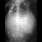
sternal cleft

Paget disease
(bone) • Paget disease of the sternum - Ganzer Fall bei Radiopaedia

Primary
closure of superior partial sternal cleft in a 2-month-old girl: case report. On the chest midline, the superior part of the skin which is in front of the heart was fibrotic, and paradoxical respiratory movements were present in this area

Primary
closure of superior partial sternal cleft in a 2-month-old girl: case report. It is seen that the superior part of the sternum was undeveloped at CT. There were hypoplastic sternal bars bilaterally. Fusion defect of the superior sternum was present, and sternal bars united at the inferior

Duplication
of the ALDH1A2 gene in association with pentalogy of Cantrell: a case report. Computed tomography scan. A sagittal view from a contrast-enhanced computed tomography study demonstrating protrusion of the right ventricle through the sternal defect (a) and extra-abdominal position of the organs in the large omphalocele (b).
Sternal clefts, also called Sternum bifidum, represent vertically oriented midline congenital defects seen at the junction of the sternal bars. They occur secondary to failure or incomplete fusion of sternal segments. The sternal defects can vary from a linear fissure to larger defects with complete or partial separation of sternum.
Associations
Large sternal clefts can be associated with ectopia cordis, omphalocele, part of complex syndromes (Cantrell's pentalogy or PHACE syndrome).
Sclerotic band
Sclerotic sternal bands are usually seen in the inferior segment in the midline.Sclerotic bands are more common than clefts, though they have no clinical significance.
Siehe auch:

 Assoziationen und Differentialdiagnosen zu Fissura sterni congenita:


