subgaleal haematoma

Cerebral
hemorrhagic contusion • Extensive subgaleal hematoma - Ganzer Fall bei Radiopaedia
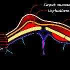
Diagram of
the infant scalp showing the locations of the common hematomata of the scalp in relation the layers of the scalp. Newborn Scalp Hematomata

Extradural
hemorrhage • Extradural hematoma, subdural hematomas, cerebral contusions and subgaleal hematoma - Ganzer Fall bei Radiopaedia

Subgaleal
hematoma • Acute tentorium cerebelli subdural hematoma - Ganzer Fall bei Radiopaedia
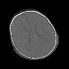
Infant
dropped by parent. Axial CT without contrast of the brain shows a left parietal depressed skull fracture with high density material in the adjacent subgaleal space that crosses sutures and courses around to the right side of the skull.The diagnosis was a depressed skull fracture causing a subgaleal hematoma.

Toddler who
is unresponsiveAxial CT without contrast of the brain shows high density material in the subgaleal tissues posteriorly, a wide lucency in the right posterior skull along with two areas of depressed lucency in the left frontal skull, a rounded high-density lesion in the midline of the cerebellum, and decreased density of the cerebrum when compared to the normal density of the cerebellum along with loss of the normal gray matter-white matter differentiation.The diagnosis was a posterior subgaleal hematoma, a right posterior diastatic skull fracture and a left frontal depressed skull fracture, cerebellar contusion and diffuse cerebral edema in a child abuse patient.

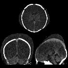
Newborn with
failure to progress during delivery due to dystocia who is now apneic. Axial CT without contrast of the brain shows a cresenteric high-density fluid collection in the subcutaneous tissues of the right scalp that crosses suture lines and a cresenteric low-density fluid collection in the subcutaneous tissues of the left scalp that crosses suture lines. Intracranially, there is diffuse loss of gray matter-white matter differentiation secondary to diffuse cerebral edema.The diagnosis was a left caput succedaneum and a right subgaleal hematoma in a patient with hypoxic ischemic encephalopathy.

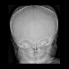
Newborn with
scalp swelling and bruising and decreased hemoglobin after a delivery by C-section for failure to descend. AP and lateral radiographs of the skull show diffuse soft tissue swelling of the scalp that crosses sutures and that is especially prominent over the left fronto-parietal region.The diagnosis was subgaleal hematoma.

Scalp •
Scalp hematoma types (diagram) - Ganzer Fall bei Radiopaedia

Subgaleal
hematoma • Parietal fracture with large subgaleal hematoma - Ganzer Fall bei Radiopaedia

Subgaleal
hematoma • Subgaleal hematoma - Ganzer Fall bei Radiopaedia

Subgaleal
hematoma • Subgaleal hematoma due to birth trauma - Ganzer Fall bei Radiopaedia

Cerebral
hemorrhagic contusion • Coup-contrecoup injury - Ganzer Fall bei Radiopaedia
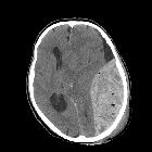
Extradural
hemorrhage • Extradural hematoma - Ganzer Fall bei Radiopaedia
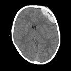
School ager
hit in head by a golf ball. Axial CT without contrast of the brain shows a high density fluid collection in the left frontal subcutaneous tissue. There is also an underlying lentiform mixed density intracranial fluid collection.The diagnosis was a left frontal subgaleal hematoma with an underlying epidural hematoma.

Subarachnoid
hemorrhage • Traumatic subarachnoid hemorrhage - coup-contrecoup injury - Ganzer Fall bei Radiopaedia
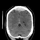
Extradural
hemorrhage • Extradural hemorrhage - Ganzer Fall bei Radiopaedia

Extradural
hemorrhage • Extradural hemorrhage with subgaleal hematoma - Ganzer Fall bei Radiopaedia
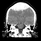
Extradural
hemorrhage • Extradural hematoma - venous - Ganzer Fall bei Radiopaedia

Extradural
hemorrhage • Extradural hemorrhage - Ganzer Fall bei Radiopaedia

Subgaleal
hematoma • Large recurrent subgaleal hematoma - Ganzer Fall bei Radiopaedia

Subgaleal
hematoma • Large traumatic subgaleal hematoma and cervical spine fracture - Ganzer Fall bei Radiopaedia

Subgaleal
hematoma • Subgaleal hematoma with retrobulbar extension - Ganzer Fall bei Radiopaedia

Subgaleal
hematoma • Subgaleal hematoma with orbital extension - Ganzer Fall bei Radiopaedia

Subgaleal
hematoma • Subgaleal hematoma with orbital extension - Ganzer Fall bei Radiopaedia

Newborn who
underwent vacuum extraction during a difficult delivery. Axial (left) and coronal (right) CT without contrast of the brain shows a small, round, dense lesion in the left cerebellar hemisphere. The coronal image also shows a dense fluid collection in the left superior subcutaneous tissues of the skull which crosses the sagittal suture in the midline.The diagnosis was left cerebellar hemorrhage and left subgaleal hematoma.
subgaleal haematoma
Siehe auch:
und weiter:

 Assoziationen und Differentialdiagnosen zu subgaleal haematoma:

