Supradiaphragmatic liver

Unusual
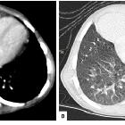
association of parvo-virus with Morgagni hernia, mistaken for patch of consolidation. Axial mediastinal (A) and lung (B) window showed a homogenous soft tissue density in RT cardio-phrenic angle abutting and displacing the pericardium and RT ventricle with clear interface. Sharp delineation with lung parenchyma is noted.

Unusual
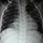
association of parvo-virus with Morgagni hernia, mistaken for patch of consolidation. Chest X-ray PA view shows radio-opacity involving the RT cardio-phrenic angle partially silhouetting the RT cardiac border. The radio-opacity showed sharp interface with adjacent lung parenchyma.

Unusual
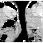
association of parvo-virus with Morgagni hernia, mistaken for patch of consolidation. Coronal (A) and Sagittal (B) reformatted images showed herniation of LT lobe of liver along with vasculature into the RT sterno-costal space with shifting of heart towards LT side.
Supradiaphragmatic liver has been reported as a very rare variant in liver morphology.
In this variant, liver tissue extends into the right hemithorax through an opening in the right hemidiaphragm. The tissue is connected to the right hepatic lobe by a pedicle. In one report, the caudate lobe herniated into the thorax .
In some cases, hepatopulmonary fusion has been described and is presumably the result of right-sided congenital diaphragmatic herniation.
The patient should not have a history of trauma, otherwise traumatic liver herniation through a diaphragmatic tear would be much more likely.
Differential diagnosis
- congenital diaphragmatic hernia
- diaphragmatic rupture: usually has a history of trauma (although may be remote)
Siehe auch:

 Assoziationen und Differentialdiagnosen zu supradiaphragmale Leber: