Swallow tail appearance


The swallow tail sign describes the normal axial imaging appearance of nigrosome-1 within the substantia nigra on high-resolution T2*/SWI weighted MRI.
Terminology
Yes, this is one of those annoying signs where its presence is normal. A normal-appearing substantia nigra is reminiscent of a swallow's tail. If nigrosome-1 loses its high signal on SWI, the split-tail appearance is lost and this should, therefore, be reported as an "absent swallow tail sign". Better yet, one can simply report the fact that nigrosome-1 cannot be visualized.
Pathology
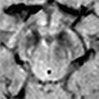
Nigrosome-1, within the posterior third of the substantia nigra, returns high signal on axial SWI in a linear or comma shape. It is surrounded anteriorly, laterally and medially by low SWI signal intensity which resembles the distinctive split tail of a swallow.
In Parkinson disease the high SWI signal within nigrosome-1 is absent and hence the swallow tail sign is absent. This is believed to be the result of abnormal iron metabolism, and two proposed mechanisms for this: increased iron content or decreased neuromelanin content with decreased iron storage capacity leading to more free iron with paramagnetic properties .
Radiographic features
MRI
It is important to note that adequate visualization of nigrosome-1 requires targeted 3D high-resolution SWI. In the original publication the parameters were:
- FEEPI, TR/TE 60/30, echo train length 5, flip angle 19°
- number of slices: 70
- voxel size 0.55 x 0.55 x 0.7 mm,
Interpretation
Absence of the sign (absent swallow tail sign) is reported to have a diagnostic accuracy of greater than 90% for Parkinson disease and dementia with Lewy bodies . In the original description, the negative predictive value (in other words visualizing nigrosome-1 bilaterally predicts the absence of Parkinson disease) was 100% and for dementia with Lewy bodies 89% .
Siehe auch:


 Assoziationen und Differentialdiagnosen zu Schwalbenschwanzzeichen: