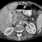
Thrombose der Arteria splenica

Imaging
findings of splenic emergencies: a pictorial review. Splenic artery thrombosis. (a) Axial contrast-enhanced arterial phase CT of a 74-year-old man with atrial fibrillation demonstrates patent (arrow) and thrombosed (arrowhead) portions of the splenic artery. (b) MIP and (c) VR CT images more clearly demonstrate thrombosed splenic (arrowhead) and patent hepatic (arrow) arteries. (d) Axial and (e) coronal contrast-enhanced CT images at venous phase reveal diffuse splenic infarct (asterisks) resulting from splenic artery thrombosis. Capsular enhancement is seen around the spleen (arrows in d)
Thrombose der Arteria splenica
Siehe auch:

 Assoziationen und Differentialdiagnosen zu Thrombose der Arteria splenica:

