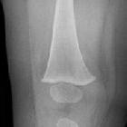
tibial hemimelia


Tibial hemimelia or tibial deficiency is an uncommon abnormality which can range from isolated mild shortening to complete tibial absence .
Epidemiology
Tibial hemimelia is extremely rare, occurring in only one out of one million live births .
Clinical presentation
The affected limb is short with varus deformity of the ankle and knee flexion contracture.
Pathology
Tibial hemimelia is usually sporadic, although an autosomal dominant inheritance pattern has been reported in bilateral cases .
Deficient bone part is usually replaced with a strong fibrous or fibrocartilaginous band .
Tibial hemimelia can be unilateral or bilateral, with 30% of cases reported bilateral. For some unknown reason, Tibial hemimelia favors the right side (72% of unilateral cases). Tibial hemimelia may also be present alongside additional congenital deformities, such as congenital femoral deficiency .
Associations
There is a high association with ipsilateral varus deformity and medial foot deficiencies . Patellar bone and quadriceps muscle may be absent.
Tibial hemimelia is associated with several syndromes:
- Werner syndrome
- tibial hemimelia-polysyndactyly-triphalangeal thumb syndrome (THPTTS)
- CHARGE syndrome
Classification
Based on the radiographic appearance, four types of tibial hemimelia have been recognized :
- type 1: absent tibia
- type 1a: with absent tibia and hypoplastic lower femoral epiphysis
- type 1b: with absent tibia but normal lower femoral epiphysis
- type 2: in which the tibia is distally deficient and well developed proximally
- type 3: in which the tibia is proximally deficient and well ossified distally
- type 4: characterized by shortening of the distal tibia, with distal tibiofibular diastasis and normally developed proximal tibia.
Radiographic features
Plain radiograph
Radiography enables excellent assessment of tibial hemimelia and its associated skeletal malformations;
- tibia may be shortened, dysplastic, or absent. The tibia may also present as a fibrous remnant that does not appear on x-rays.
- the fibula is normal or dysplastic. It is often dislocated from the knee.
- foot deformities are present as well.
Differential diagnosis
- fibular hemimelia: in tibial hemimelia, the foot and ankle are always in varus position (pointing inward) while in fibular hemimelia they are almost always in valgus position (pointing outward)
- congenital deficiency of the knee: both of these conditions demonstrate congenital deficiency of the tibia and relative fibular overgrowth
- proximal femoral focal deficiency
See also
Siehe auch:
- Polydaktylie
- Kryptorchismus
- Syndaktylie
- Fibulare Hemimelie
- Hemimelie
- Eaton-McKusick-Syndrom
- Femurhypoplasie
und weiter:

 Assoziationen und Differentialdiagnosen zu Tibiale Hemimelie:


