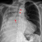
TNM staging of lung cancer
The IASLC (International Association for the Study of Lung Cancer) 8 edition lung cancer staging system was introduced in 2016 and supersedes the IASLC 7 edition.
Standard-of-care lung cancer staging ideally should be performed in a multidisciplinary meeting using the information provided both from CT and FDG-PET/CT with further inputs from the histopathologic findings (pathological staging). The National Comprehensive Cancer Network (NCCN) guidelines recommend that FDG-PET/CT should be offered to all patients with non-small cell lung cancer (NSCLC) and that PET-positive findings for mediastinal nodes and/or distant disease require histopathological or other radiological confirmation .
TNM system
T: primary tumor
- Tx: primary tumor cannot be assessed or tumor proven by the presence of malignant cells in sputum or bronchial washings but not visualized by imaging or bronchoscopy
- T0: no evidence of a primary tumor
- Tis: carcinoma in situ - tumor measuring 3 cm or less and has no invasive component at histopathology
- T1: tumor measuring 3 cm or less in greatest dimension surrounded by lung or visceral pleura without bronchoscopic evidence of invasion more proximal than the lobar bronchus (i.e. not in the main bronchus)
- T1a(mi): minimally invasive adenocarcinoma
- tumor has an invasive component measuring 5 mm or less at histopathology
- T1a ss: superficial spreading tumor in central airways (spreading tumor of any size but confined to the tracheal or bronchial wall)
- T1a: tumor ≤1 cm in greatest dimension
- T1b: tumor >1 cm but ≤2 cm in greatest dimension
- T1c: tumor >2 cm but ≤3 cm in greatest dimension
- T1a(mi): minimally invasive adenocarcinoma
- T2: tumor >3 cm but ≤5 cm or tumor with any of the following features:
- involves the main bronchus regardless of distance from the carina but without the involvement of the carina
- invades visceral pleura
- associated with atelectasis or obstructive pneumonitis that extends to the hilar region
- involving part or all of the lung
- T2a: tumor >3 cm but ≤4 cm in greatest dimension
- T2b: tumor >4 cm but ≤5 cm in greatest dimension
- T3: tumor >5 cm but ≤7 cm in greatest dimension or associated with separate tumor nodule(s) in the same lobe as the primary tumor or directly invades any of the following structures:
- chest wall (including the parietal pleura and superior sulcus)
- phrenic nerve
- parietal pericardium
- T4:
- tumor >7 cm in greatest dimension or associated with separate tumor nodule(s) in a different ipsilateral lobe than that of the primary tumor or
- invades any of the following structures
- diaphragm
- mediastinum
- heart
- great vessels
- trachea
- recurrent laryngeal nerve
- esophagus
- vertebral body
- carina
It is recommended that solid and non-solid lesions should be measured on the image that shows the greatest tumor dimension (on axial, coronal, or sagittal planes). Although those lesions that are part solid should be measured on both their largest average diameter and the largest diameter of the solid component, only the solid component measurement is to be used for staging directions . Also, the solid component of subsolid lesions should be performed on a lung or intermediate window rather than mediastinal window .
For those centrally located lung tumors associated with peripheral post-obstructive atelectasis, FDG-PET/CT is useful in further delineating the tumor real size and, therefore, leads to a more precise T staging and, if it is the case, to a smaller targeted volume in radiation treatment planning.
N: regional lymph node involvement
- Nx: regional lymph nodes cannot be assessed
- N0: no regional lymph node metastasis
- N1: metastasis in ipsilateral peribronchial and/or ipsilateral hilar lymph nodes and intrapulmonary nodes, including involvement by direct extension
- N2: metastasis in ipsilateral mediastinal and/or subcarinal lymph node(s)
- N3: metastasis in contralateral mediastinal, contralateral hilar, ipsilateral or contralateral scalene, or supraclavicular lymph node(s)
Please note that there has been no change in nodal involvement staging since the 7 edition of the IASLC.
PET-CT plays an important role in staging nodal disease. FDG uptake higher than the blood pool is suspicious, and uptake higher than the liver it is highly concerning for nodal metastases. Endobronchial biopsy of an FDG-avid node is recommended to confirm the highest pathologic stage of disease .
M: distant metastasis
- M0: no distant metastasis
- M1: distant metastasis present
- M1a: separate tumor nodule(s) in a contralateral lobe; tumor with pleural or pericardial nodule(s) or malignant pleural or pericardial effusions
- M1b: single extrathoracic metastasis, involving a single organ or a single distant (nonregional) node
- a single extrathoracic metastasis has a better survival and different treatment choices, which is why it has now been staged separately
- M1c: multiple extrathoracic metastases in one or more organs
NB: The MX category is no longer used, it was removed in the 6 edition of the TNM system, if presence of metastases is not known the cancer is assigned M0 .
There is a recommendation that the number of metastatic lesions, the larger diameter of individual metastatic deposits, and the number of involved organs should be stated in the radiological report . However, note that the site of the metastasis by itself is not a prognostic factor .
FDG PET/CT has a higher diagnostic value for the diagnosis of bone metastases compared to other methods. Therefore, bone scintigraphy is not recommended for staging purposes .
Histologic diagnosis is recommended when the adrenal gland is the only site of metastatic disease, given the risk of a false-positive .
Stage groupings
- stage 0
- TNM equivalent: Tis, N0, M0
- stage Ia
- TNM equivalent: T1, N0, M0
- 5-year survival: up to 92%
- stage Ib
- TNM equivalent: T2a, N0, M0
- 5-year survival: 68%
- stage IIa
- TNM equivalent: T2b, N0, M0
- 5-year survival: 60%
- stage IIb
- TNM equivalent: T1/T2, N1, M0 or T3, N0, M0
- 5-year survival: 53%
- stage IIIa
- TNM equivalent: T1/T2, N2, M0 or T3/T4, N1, M0 or T4, N0, M0
- 5-year survival: 36%
- stage IIIb
- TNM equivalent: T1/T2, N3, M0 or T3/T4, N2, M0
- 5-year survival: 26%
- stage IIIc
- TNM equivalent: T3/T4, N3, M0
- 5-year survival: 13%
- stage IVa
- TNM equivalent: any T, any N with M1a/M1b
- 5-year survival: 10%
- stage IVb
- TNM equivalent: any T, any N with M1c
- 5-year survival: 0%
Siehe auch:
und weiter:

 Assoziationen und Differentialdiagnosen zu Lungenkarzinom Staging:

