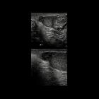
torsion of the appendix testis

Preschooler
with acute onset of left scrotal pain. Transverse (above) and sagittal (below) US of the left testicle shows a round, mixed echogenicity lesion superior and medial to the left testicle that had no flow on color and spectral doppler US. The left epididymis and left testicle were normal in appearance with normal appearing blood flow.The diagnosis was torsion of the appendix testis on the left.

Torsion of
the appendix testis • Torsion of the testicular appendix - Ganzer Fall bei Radiopaedia

Torsion of
the appendix testis • Torsion of the hydatid of Morgagni / testicular appendix - Ganzer Fall bei Radiopaedia

Torsion of
the appendix testis • Torsion of Morgagni appendix - Ganzer Fall bei Radiopaedia

Torsion of
appendix testis with blue dot sign. Longitudinal views sonogram showing a hypoechogenic, well-define nodule (between calipers) between the upper pole of left testis (T) and epididymal head (E), related to twisted hydatid of Morgagni (H).

Torsion of
appendix testis with blue dot sign. Longitudinal view Colour Doppler sonogram reveals that the nodule is avascular (arrows) and the tissue rounded the nodule is hypervascularized.

Torsion of
the appendix testis • Torsion of the appendix testis - Ganzer Fall bei Radiopaedia

Torsion of
appendix testis with blue dot sign. Physical examination showed a blue dot sign (arrow) and a congestive vein (arrowheads) in the upper left scrotum pole.
Torsion of the appendix testis (occasionally called torsion of the hydatid of Morgagni) is the most common cause of an acute painful hemiscrotum in a child. The appendix testis is located at the upper pole of the testis (between the testis and the head of the epididymis).
The normal appendix testis is 1 to 4 mm in length, and it is oval or pedunculated in shape.
Clinical presentation
Blue dot sign:
- classic finding on physical examination
- small firm nodule is palpable on the superior aspect of the testis and exhibits bluish discoloration through the overlying skin
Radiographic features
Ultrasound
Appendix testis is increased in size with an increase or decrease in echogenicity. Torsion of the appendix testis is frequently accompanied by hydrocele and scrotal wall thickening .
A spherical shape and size of 6 mm with no internal vascularity and peripheral vascularity on Doppler scan are highly suggestive of torsion.
See also
Siehe auch:
und weiter:

 Assoziationen und Differentialdiagnosen zu Hydatidentorsion:


