Trachealstenose


Tracheal stenosis is usually acquired following intubation or tracheostomy. It can also arise as part of the spectrum of tracheobronchial stenosis.
Pathology
Inflammation and pressure necrosis of the tracheal mucosa most commonly occur at either the tracheostomy stoma or at the level of the tube balloon. Acute post-intubation stenosis results from mucosal edema or granulation tissue.
The stenosis is typically 1.5-2.5 cm in length. In patients with chronic stricture, tracheomalacia may result from weakness of tracheal cartilage and can be a cause of dyspnea.
Radiographic features
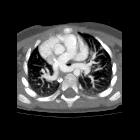
CT
Eccentric or concentric soft tissue thickening internal to normal-appearing tracheal cartilage may be visible. The outer tracheal wall has a normal appearance without evidence of deformity or narrowing. Expiratory CT shows little change in tracheal diameter.
Siehe auch:
- saber-sheath trachea
- Rezidivierende Polychondritis
- Trachealkompression durch den Truncus brachiocephalicus
- diffuse Trachealverengung
- Trachealstenose bei Kindern
und weiter:


 Assoziationen und Differentialdiagnosen zu Trachealstenose:

