tuberkulöse Spondylodiscitis

Extrapulmonary
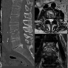
tuberculosıs: an old but resurgent problem. MR images of lumbar vertebrae of a 47-year-old male with tuberculous spondylodiscitis. Sagittal T2-weighted image (a) shows the heterogeneous low signal in the anterior parts of the L3 and L4 vertebral bodies and L3-4 intervertebral disc, which offers minimal anterior subligamentous extension (arrows). Pre-contrast (a) and post-contrast (b) T1 weighted images reveal diffuse contrast enhancement of both vertebral bodies in keeping with spondylodiscitis (arrowheads)

Urinary
tuberculosis: multidetector CT findings. Sagittal T1-(a), STIR (b) and T2-weighted (c) images inflammatory-type signal changes (*) affecting the L4 and L5 vertebral bodies and fluid content in the intervertebral disk (arrows in b&c) suggesting infectious spondylo-diskitis.

Urinary
tuberculosis: multidetector CT findings. Sagittal T1-(a), STIR (b) and T2-weighted (c) images inflammatory-type signal changes (*) affecting the L4 and L5 vertebral bodies and fluid content in the intervertebral disk (arrows in b&c) suggesting infectious spondylo-diskitis.

Urinary
tuberculosis: multidetector CT findings. Sagittal T1-(a), STIR (b) and T2-weighted (c) images inflammatory-type signal changes (*) affecting the L4 and L5 vertebral bodies and fluid content in the intervertebral disk (arrows in b&c) suggesting infectious spondylo-diskitis.

Urinary
tuberculosis: multidetector CT findings. Axial (d) and coronal (e,f) T2-weighted image revealed bilateral paravertebral abscesses (+), associated with fluid content in the intervertebral disk (arrow) suggesting tuberculous spondylo-diskitis.

Urinary
tuberculosis: multidetector CT findings. Note paravertebral abscesses (+), liquefied L4-L4 intervertebral disk (arrow). Additionally, moderate left-sided hydronephrosis (*), right kidney with parenchymal thinning, dilated and distorted calyces (thin arrows), non-dilated pelvis with mild mural thickening (thick arrow) were noted.

Urinary
tuberculosis: multidetector CT findings. A sagittal CT reformatted image viewed at bone window settings showed destructive vertebral changes (*) corresponding to the MR signal changes in the L4 and L5 vertebral bodies.

Urinary
tuberculosis: multidetector CT findings. Detail nephrographic phase images better showed atrophied right kidney with thinned parenchyma and delayed enhancement, uneven caliectasis (thin arrows). Note left hydronephrosis (*), paraspinal abscesses (+).

Urinary
tuberculosis: multidetector CT findings. Detail nephrographic phase images better showed mild enhancing urothelial thickening (thick arrows) along the right ureter, and bilateral paraspinal abscesses (+).
tuberkulöse Spondylodiscitis
Siehe auch:
und weiter:

 Assoziationen und Differentialdiagnosen zu tuberkulöse Spondylodiscitis:

