urethral stricture


 nicht verwechseln mit: Urethralklappe
Urethral strictures are relatively common and typically occur either in the setting of trauma or infection.
Epidemiology
The demographics of the affected population is dictated by the etiology, but in general, it is safe to say that adult males make up the vast majority of cases.
Clinical presentation
The primary mode of presentation of a symptomatic stricture is with poor urinary stream. If severe, bladder hypertrophy and trabeculation may occur.
Pathology
In general, the term urethral stricture refers to a fibrous scarring of the anterior urethra caused by collagen and fibroblast proliferation.
Etiology
Common causes of urethral strictures include:
- infection
- gonococcal urethritis (more common)
- non-gonococcal urethritis (less common)
- inflammatory
- balanitis xerotica obliterans
- trauma
- straddle injury (most common)
- pelvic fractures
- iatrogenic
- instrumentation
- prolonged catheterization
- transurethral resection of the prostate
- open radical prostatectomy
- urethra reconstruction (hypospadias/epispadias)
- congenital
- uncommon
- not to be confused with posterior urethral valves
Although gonorrhea remains the most common sexually transmitted disease, urethral strictures are far less common than previously due to early treatment.
Instrumentation-related strictures usually occur in the bulbomembranous region and, less commonly, at the penoscrotal junction.
Radiographic features
Radiographic evaluation helps define the location, length, number, and degree of strictures.
Fluoroscopy
Retrograde urethrography is the primary method used to image anterior urethral stricture.
In posterior urethral strictures following blunt trauma, simultaneous antegrade cystourethrography and retrograde urethrography are often required to determine the length of the urethral defect.
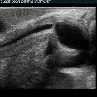
Ultrasound
Sonourethrography is best used adjunctively to guide treatment planning in patients with known bulbous urethral strictures and has been reported to be more accurate than retrograde urethrography for estimating the length of urethral strictures.
In pediatric cases, it worth using ultrasound instead of x-rays and CT to reduce dose to the patient, although this may be a challenging test due to lack of cooperation.
MRI
MR imaging is considered to be the best ancillary imaging modality for assessing post-traumatic pelvic anatomy.
Treatment and prognosis
Treatment options include urethral dilation, internal urethrotomy, or permanent urethral stenting.
Practical pearls and pitfalls
Urethral strictures should not be confused with physiologic narrowing of the lumen at the level of membranous part of the urethra.
Siehe auch:
und weiter:


 Assoziationen und Differentialdiagnosen zu Harnröhrenstriktur: