Vena portae


The portal vein (PV) (sometimes referred to as the main or hepatic portal vein) is the main vessel in the portal venous system and drains blood from the gastrointestinal tract and spleen to the liver.
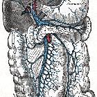
Gross anatomy
The portal vein usually measures approximately 8 cm in length in adults with a maximum diameter of 13 mm. It originates behind the neck of the pancreas where it is classically formed by the confluence of the superior mesenteric and splenic veins (the portovenous confluence), and also receives blood from the inferior mesenteric, gastric, and cystic veins.
Immediately before reaching the liver, the portal vein divides in the porta hepatis into left and right portal veins. The right portal vein divides into anterior (supplying segments 5 and 8) and posterior (supplying segments 6 and 7) branches. The left portal vein may be divided into transverse and umbilical portions, as delineated by the ligamentum venosum, and is mostly extrahepatic in course. The main branches of the left portal vein originate from the umbilical portion, and supply liver segments 2, 3 and 4 .
It ramifies further, forming smaller venous branches and ultimately portal venules. Each portal venule courses alongside a hepatic arteriole and the two vessels form the vascular components of the portal triad. These vessels ultimately empty into the hepatic sinusoids to supply blood to the liver.
75% of the blood supplied to the liver comes from the portal vein, but it only supplies 50% of the oxygen supply to the liver.
Relations
- Anterior - proper hepatic artery, the neck of pancreas, D1 segment of duodenum
- Anterolateral - common bile duct
- Posterior - inferior vena cava
Variant anatomy
The overall incidence of portal vein variation is reported to be ~25% (range 20-30%), which should be recognized prior to procedures such as liver transplantation, complex hepatectomy and portal vein embolization :
- portal vein trifurcation (most common)
- portal vein divides into three branches: left portal vein, right anterior portal vein, and right posterior portal vein
- absent right portal vein (rare)
- right sectional portal veins originate independently from the common portal vein
- if the right anterior section portal vein branches higher from the common portal vein vs posterior sectional portal vein, the surgeon may mistake the posterior sectional portal vein for the right portal vein
- portal vein duplication (rare)
- absent left extrahepatic portal vein (rare)
- a single right portal vein originates from the porta hepatis, supplying the right hemiliver, then following an intrahepatic course with distalmost branches supplying the left liver
- preduodenal portal vein
- frequently associated with other abnormalities and situs inversus
There is an increased risk of bile duct hilar anatomical variation in the presence of portal vein variants.
Development
The embryonic vitelline veins drain from the yolk sac to the sinus venosus. The two vitelline veins develop anastomosing cross-communications around the duodenum, in the developing liver and the septum transversum. Selective involution of these veins leads to the formation of the portal vein and rotation of the foregut contributes to the formation of its extra-hepatic course .
Abnormal involution can lead to congenital abnormalities, including a congenital portosystemic shunt.
Related pathology
Siehe auch:
und weiter:


 Assoziationen und Differentialdiagnosen zu Vena portae:

