kavernöse Transformation Pfortader


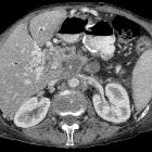
Cavernous transformation of the portal vein (CTPV) is a sequela of portal vein thrombosis and is the replacement of the normal single channel portal vein with numerous tortuous venous channels.
For a discussion of demographics and presentation, please refer to the article on portal vein thrombosis.
Pathology
Following thrombosis, the portal vein may or may not re-canalize. Re-canalization is seen more frequently in patients without cirrhosis or disease of the liver leading to inherently increased resistance to portal flow. In patients whose portal vein does not recanalize, or only partially re-canalizes, collateral veins (thought to be paracholedochal veins) dilate and become serpiginous.
This process takes a variable amount of time, from as little as a week to a year .
These vessels drain variably into the left and right portal veins or more distally into the liver. Additional communications can also be identified with the pericholecystic veins.
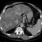
Cavernous transformation of the portal vein is most of the times inefficient in guaranteeing adequate portal vein inflow to the liver parenchyma far from the hilum and, therefore, is associated with an increased hepatic arterial flow to those peripheral liver segments. These changes lead to central liver hypertrophy and peripheral liver atrophy .
Radiographic features
In addition to direct visualization of the dilated vessels, the resultant portal hypertension results in other frequent changes: see portal hypertension. Additionally, there are changes in liver shape which are somewhat different to those seen in cirrhosis . Typically these changes are:
- atrophy of the left lateral segment (segments 2 and 3) whereas hypertrophy is more common in cirrhosis
- hypertrophy of segment 4 whereas atrophy is more common in cirrhosis
- hypertrophy of the caudate lobe which is also seen in cirrhosis
Ultrasound
Ultrasound is able to identify a normal portal vein in almost all cases when present (97%) , and therefore is a good first-order examination. Doppler examination can be carried out at the same time to evaluate for portal hypertension. Cavernous transformation appears as numerous tortuous vessels occupying the portal vein bed. Flow is generally hepatopetal and continuous with little if any respiratory or cardiac variation .
CT
Multiphase CT can confirm the diagnosis by demonstrating:
- numerous vascular structures in the region of the portal vein, which enhance during the portal venous phase, and not during the arterial phase (distinguishing it from an arteriovenous malformation)
- possible linear areas of calcification within the previously thrombosed portal vein indicating chronic venous thrombosis
MRI
MRI is also a proven method for imaging the portal venous system and may be used as a complementary or alternative modality to CT. MRI is usually reserved to clarify associated benign hepatocellular nodules that may be seen in up to a fifth of the patients, particularly the focal nodular hyperplasia-like lesions .
Treatment and prognosis
Despite collateral formation portal hypertension is usually present (up to 90%) with associated complications.
Whereas portal hypertension can in some cases be treated with TIPS, the absence of normal portal circulation usually makes this impossible.
Siehe auch:
- Leberzirrhose
- Arteriovenöse Malformation
- Hypertrophie des Lobus caudatus
- Pfortaderthrombose
- Pankreaskarzinom
- Vena portae
- hepatopetal
- portale Hypertension
- transjugulärer intrahepatischer portosystemischer Shunt
- portale Biliopathie
- segment IV
- segments 2 and 3
und weiter:

 Assoziationen und Differentialdiagnosen zu kavernöse Transformation Pfortader:


