venöse Kanäle und Lakunen der Kalotte
Venous lacunae also known as venous lakes are enlarged venous spaces within the skull, most often in the parasagittal region. They are normal variants and their primary importance is that they may mimic lytic lesions.
Anatomy
Venous lacunae are the result of focal venous dilatations located within the diploid space of the skull, most commonly encountered adjacent to the superior sagittal sinus. They receive blood from the cerebral veins as well as meningeal veins and connect to diploic and emissary veins .
They can be invaginated by arachnoid granulations and therefore act as a means of CSF absorption .
Radiographic features
Plain radiograph
Venous lacunes are seen as a lyric area adjacent to the sagittal suture, mainly in the parietal bone.
CT
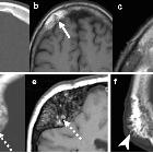
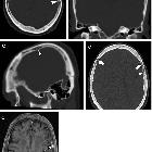
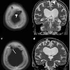
On CT, venous lacunes appear as sharply demarcated regions of lucency with blood density centrally. As they are venous structures, they demonstrate enhancement on delayed postcontrast studies .
MRI
Similarly, on MRI venous lacunes resemble other venous structures.
Siehe auch:
und weiter:


 Assoziationen und Differentialdiagnosen zu venöse Kanäle und Lakunen der Kalotte: