zerebelläres Lymphom

Cerebellar
large B-cell lymphoma: a case report. CT with contrast showing enhancing cerebellar lesion (yellow arrow) and vasogenic edema (red arrow)

Cerebellar
large B-cell lymphoma: a case report. PET scan showing cerebellar hypermetabolic lesions (orange arrow)

Clinicoradiological
changes of brain NK/T cell lymphoma manifesting pure akinesia: a case report. Brain T2-weighted imaging at neurological onset. A-F. Hyperintense lesions are observed in the dorsal pons, midbrain, cerebellum and periventricular white matter. E. There were no remarkable changes in the basal ganglia.

Clinicoradiological
changes of brain NK/T cell lymphoma manifesting pure akinesia: a case report. Brain MRI after neurological worsening. A-D. T2-weighted image show widespread T2-hyperintense lesions in the brainstem, cerebellum, basal ganglia and cerebral white matter. E-H. DWI shows hyperintense lesions in the middle cerebellar peduncles, midbrain, left internal capsules and cerebral white matter. I-L. ADC shows hypointense lesions in the middle cerebellar peduncles, midbrain, left internal capsules and cerebral white matter.

Clinicoradiological
changes of brain NK/T cell lymphoma manifesting pure akinesia: a case report. Gadolinium-enhanced T1-weighted imaging. A, E. Gadolinium enhancement are found in the cerebellum (arrows) and bilateral frontal subcortex (arrowheads). B-D, F. There is no enhancement in the brainstem and basal ganglia.
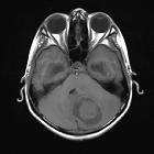
zerebelläres Lymphom
ZNS Lymphom Radiopaedia • CC-by-nc-sa 3.0 • de
CNS lymphoma refers to the involvement of the central nervous system with lymphoma. It can be broadly divided into primary and secondary, with a number of special types of also recognized.
- primary CNS lymphoma (PCNSL)
- secondary CNS lymphoma
- parenchymal
- leptomeningeal
Note that this is a simplified version of the current WHO classification of CNS tumors, which divides CNS lymphoma into a larger number of subtypes.
Siehe auch:


 Assoziationen und Differentialdiagnosen zu zerebelläres Lymphom: