zervikale venöse Malformationen

Neck mass -
the power of radiology to avoid biopsy when it is not necessary!. US shows a hypoechoic lesion with hyperechoic linear striations, without apparent vascular flow on colour Doppler.

Neck mass -
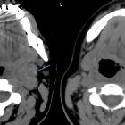
the power of radiology to avoid biopsy when it is not necessary!. Axial non-contrast CT images show subtle hyperdense foci (arrow in a) within a left level II soft tissue mass and nonspecific surrounding fatty "inflammatory" change (arrow in b).

Neck mass -
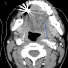
the power of radiology to avoid biopsy when it is not necessary!. Multiphase axial CT images with contrast show a trans-spatial lesion that enhances slowly and demonstrates some venous wash-out.

Neck mass -
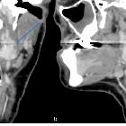
the power of radiology to avoid biopsy when it is not necessary!. Coronal (a) and sagittal (b) CT images demonstrate the venous malformation.

Neck mass -
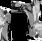
the power of radiology to avoid biopsy when it is not necessary!. Curved multiplanar reformatted images better depicte the extension of the lesion.

Neck mass -
the power of radiology to avoid biopsy when it is not necessary!. Axial (a) and coronal (b) STIR images show a hyperintense vascular malformation.

Neck mass -
the power of radiology to avoid biopsy when it is not necessary!. T1 (a) and T2 (b) axial MR images show an isointense to muscle (T1) and hyperintense (T2) lesion abutting the oropharyngeal wall.

Neck mass -
the power of radiology to avoid biopsy when it is not necessary!. MR image with gadolinium shows strong internal enhancement.
zervikale venöse Malformationen
Siehe auch:

 Assoziationen und Differentialdiagnosen zu zervikale venöse Malformationen:


