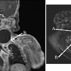
zervikales zystisches Lymphangiom


zervikales zystisches Lymphangiom
zystisches Lymphangiom Radiopaedia • CC-by-nc-sa 3.0 • de
Cystic hygroma, also known as cystic or nuchal lymphangioma, refers to the congenital macrocystic lymphatic malformations that most commonly occur in the cervicofacial regions, particularly at the posterior cervical triangle in infants.
Terminology
While these lesions are commonly known as cystic hygromas or cystic lymphangiomas, the most up-to-date terminology from ISSVA refers to them as macrocystic lymphatic malformations .
Epidemiology
They usually occur in the fetal/infantile and pediatric populations with most lesions presenting by the age of two. The estimated prevalence in the fetal population is 0.2-3%.
Clinical presentation
Patients in the infantile or pediatric population can present with pain, dyspnea, infection, hemorrhage, or respiratory compromise.
Pathology
They are thought to arise from delayed development/maldevelopment/failure of the lymphatic system to communicate with the venous system of the neck. Like other lymphangiomas, they are endothelial lined cavernous lymphatic spaces.
Microscopically, they are comprised of endothelium-lined cystic spaces with scanty stroma. They can vary significantly in size. Lymphatic vascular malformations may be mixed with other forms of vascular malformation, including capillary or venous.
Location
They occur most commonly in the neck, which is then also termed nuchal cystic hygroma (occurs in ~80% of cases) and axilla, with only 10% of cases extending to the mediastinum and only 1% confined to the chest .
Associations
Associated anomalies can be common:
- aneuploidic anomalies: ~65% (range 50-80%) of cystic hygromas can be associated with an aneuploidic abnormality
- Turner syndrome: most frequent aneuploidic association
- Down syndrome: second most common aneuploidic association
- trisomy 13
- trisomy 18
- triploidy
- non-aneuploidic
- congenital cardiac anomalies
- aortic coarctation: most common cardiovascular anomaly
- hypoplastic left heart syndrome
- pentalogy of Cantrell
- Apert syndrome
- Cornelia de Lange syndrome
- fetal alcohol syndrome
- Fryns syndrome
- lethal multiple pterygium syndrome
- limb hypertrophy
- Noonan syndrome
- Pena Shokeir syndrome
- congenital cardiac anomalies
Radiographic features
They are usually well-circumscribed and are of fluid density. Cystic hygromas may also have an infiltrative appearance and may be uni or multilocular. The density can also be variable with a combination of fluid, soft-tissue density and fat.
Antenatal ultrasound
On prenatal ultrasound, they may present as a nuchal cyst and may show septations +/- evidence of fetal anasarca/hydrops fetalis. The presence of septations may indicate a poorer outcome. Greater volumes (>75 mm according to one study ) are thought to correlate with increased karyotypic abnormality and more unfortunate fetal outcome .
Compared to nuchal translucency, It has a higher risk for aneuploidy (5-fold), cardiac anomalies (12-fold) and fetal demise (6-fold) .
CT
Commonly seen as a hypoattenuating ill-defined neck cystic mass.
MRI
Reported signal characteristics include:
- T1: predominantly low signal unless there are hemorrhagic components
- T2: predominantly high signal
- T1C+ (Gd): no enhancement on any component except occasional faint enhancement of rim
Treatment and prognosis
Management may be by surgical excision or by injection with OK-432, a preparation containing Streptococcus pyogenes antigens, which induces an inflammatory response and subsequent obliteration of the abnormal cavities. Most fetuses with cystic hygromas have a poor prognosis although it may improve in utero on its own in a tiny proportion of cases. Spontaneous remission does not necessarily exclude an abnormal karyotype.
Complications
- development of non-immune hydrops fetalis: which often indicates a poorer prognosis
- respiratory obstruction from pharyngeal edema
Differential diagnosis
Differential considerations on antenatal ultrasound include:
See also
Siehe auch:
- zystische Lymphknoten
- Meningozele
- Lymphangiom
- zystisches Lymphangiom
- zervikale bronchogene Zyste
- zystisches Lymphangiom des Rückens
- retroperitoneales Lymphangiom
- kongenitales zervikales Teratom
- Lymphozele
- occipital encephalocoele
- Serom
und weiter:

 Assoziationen und Differentialdiagnosen zu zervikales zystisches Lymphangiom:


